

Mesenchymal Stromal Cell-Derived Factors Promote Tissue Repair in a Small-for-Size Ischemic Liver Model but Do Not Protect against Early Effects of Ischemia and Reperfusion Injury
- PMID: 26380314
- PMCID: PMC4561317
- DOI: 10.1155/2015/202975
Mesenchymal Stromal Cell-Derived Factors Promote Tissue Repair in a Small-for-Size Ischemic Liver Model but Do Not Protect against Early Effects of Ischemia and Reperfusion Injury
Abstract
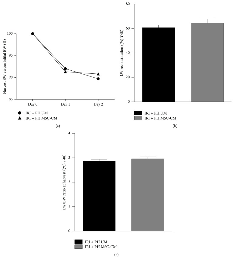
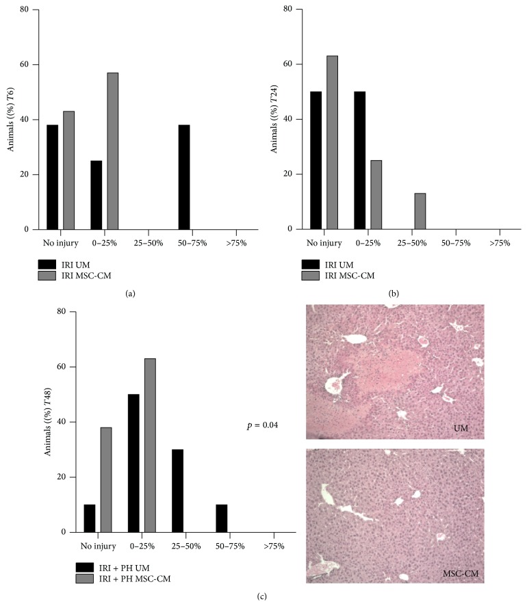
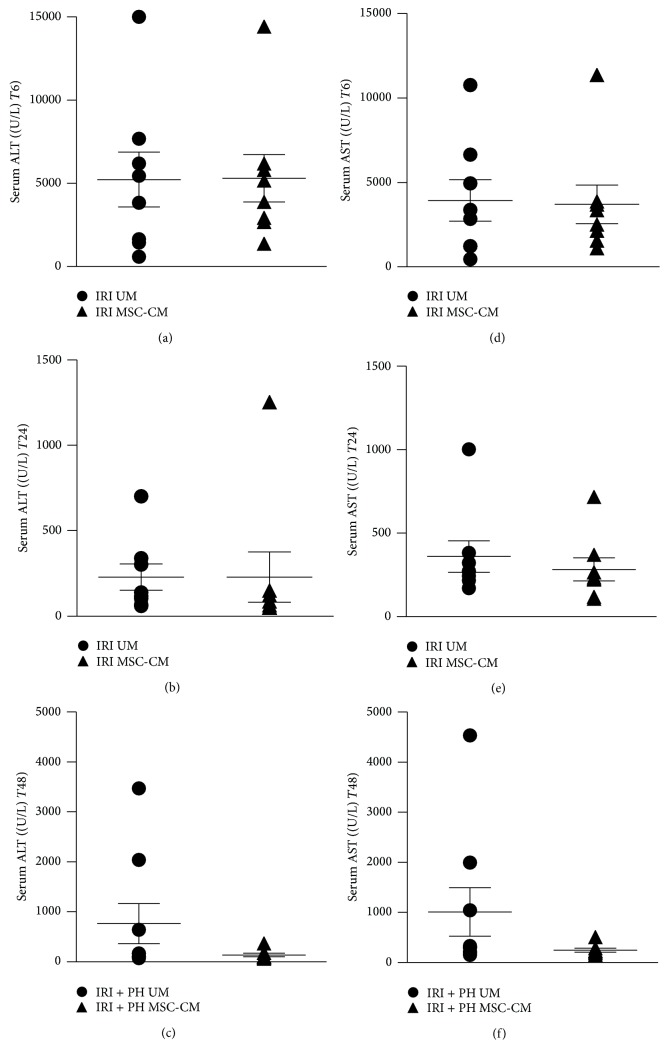
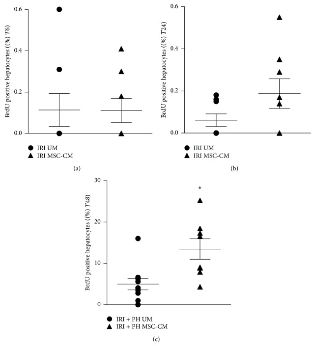
Loss of liver mass and ischemia/reperfusion injury (IRI) are major contributors to postresectional liver failure and small-for-size syndrome. Mesenchymal stromal cell- (MSC-) secreted factors are described to stimulate regeneration after partial hepatectomy. This study investigates if liver-derived MSC-secreted factors also promote liver regeneration after resection in the presence of IRI. C57BL/6 mice underwent IRI of 70% of their liver mass, alone or combined with 50% partial hepatectomy (PH). Mice were treated with MSC-conditioned medium (MSC-CM) or unconditioned medium (UM) and sacrificed after 6 or 24 hours (IRI group) or after 48 hours (IRI + PH group). Blood and liver tissue were analyzed for tissue injury, hepatocyte proliferation, and gene expression. In the IRI alone model, serum ALT and AST levels, hepatic tissue damage, and inflammatory cytokine gene expression showed no significant differences between both treatment groups. In the IRI + PH model, significant reduction in hepatic tissue damage as well as a significant increase in hepatocyte proliferation was observed after MSC-CM treatment.
Conclusion: Mesenchymal stromal cell-derived factors promote tissue regeneration of small-for-size livers exposed to ischemic conditions but do not protect against early ischemia and reperfusion injury itself. MSC-derived factors therefore represent a promising treatment strategy for small-for-size syndrome and postresectional liver failure.
Figures


Similar articles
-
Secreted factors of human liver-derived mesenchymal stem cells promote liver regeneration early after partial hepatectomy.Stem Cells Dev. 2012 Sep 1;21(13):2410-9. doi: 10.1089/scd.2011.0560. Epub 2012 May 15. Stem Cells Dev. 2012. PMID: 22455365
-
Human Mesenchymal Stromal Cell-Derived Extracellular Vesicles Improve Liver Regeneration After Ischemia Reperfusion Injury in Mice.Stem Cells Dev. 2019 Nov 1;28(21):1451-1462. doi: 10.1089/scd.2019.0085. Epub 2019 Oct 8. Stem Cells Dev. 2019. PMID: 31495270
-
Mesenchymal stem cell-conditioned medium reduces liver injury and enhances regeneration in reduced-size rat liver transplantation.J Surg Res. 2013 Aug;183(2):907-15. doi: 10.1016/j.jss.2013.02.009. Epub 2013 Mar 6. J Surg Res. 2013. PMID: 23522455
-
Mesenchymal Stromal Cell Therapy in Ischemia/Reperfusion Injury.J Immunol Res. 2015;2015:602597. doi: 10.1155/2015/602597. Epub 2015 Jul 15. J Immunol Res. 2015. PMID: 26258151 Free PMC article. Review.
-
Mesenchymal Stem Cells in Renal Ischemia-Reperfusion Injury: Biological and Therapeutic Perspectives.Curr Stem Cell Res Ther. 2017;12(3):183-187. doi: 10.2174/1574888X11666161024143640. Curr Stem Cell Res Ther. 2017. PMID: 27781940 Review.
Cited by
-
PPAR-gamma activation is associated with reduced liver ischemia-reperfusion injury and altered tissue-resident macrophages polarization in a mouse model.PLoS One. 2018 Apr 4;13(4):e0195212. doi: 10.1371/journal.pone.0195212. eCollection 2018. PLoS One. 2018. PMID: 29617419 Free PMC article.
-
Spheroid-cultured human umbilical cord-derived mesenchymal stem cells attenuate hepatic ischemia-reperfusion injury in rats.Sci Rep. 2018 Feb 6;8(1):2518. doi: 10.1038/s41598-018-20975-0. Sci Rep. 2018. PMID: 29410537 Free PMC article.
-
Mesenchymal Stem Cells Transplantation following Partial Hepatectomy: A New Concept to Promote Liver Regeneration-Systematic Review of the Literature Focused on Experimental Studies in Rodent Models.Stem Cells Int. 2017;2017:7567958. doi: 10.1155/2017/7567958. Epub 2017 Mar 13. Stem Cells Int. 2017. PMID: 28386285 Free PMC article. Review.
-
Adipose-Derived Stem Cell Transplantation Attenuates Inflammation and Promotes Liver Regeneration after Ischemia-Reperfusion and Hemihepatectomy in Swine.Stem Cells Int. 2019 Nov 18;2019:2489584. doi: 10.1155/2019/2489584. eCollection 2019. Stem Cells Int. 2019. PMID: 31827526 Free PMC article.
-
Protective effects of heme oxygenase-1-transduced bone marrow-derived mesenchymal stem cells on reduced‑size liver transplantation: Role of autophagy regulated by the ERK/mTOR signaling pathway.Int J Mol Med. 2017 Nov;40(5):1537-1548. doi: 10.3892/ijmm.2017.3121. Epub 2017 Sep 6. Int J Mol Med. 2017. PMID: 28901391 Free PMC article.
References
Publication types
MeSH terms
Substances
LinkOut - more resources
Full Text Sources
Other Literature Sources
