

Emerging and Evolving Ovarian Cancer Animal Models
- PMID: 26380555
- PMCID: PMC4558890
- DOI: 10.4137/CGM.S21221
Emerging and Evolving Ovarian Cancer Animal Models
Abstract
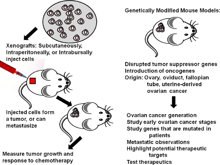
Ovarian cancer (OC) is the leading cause of death from a gynecological malignancy in the United States. By the time a woman is diagnosed with OC, the tumor has usually metastasized. Mouse models that are used to recapitulate different aspects of human OC have been evolving for nearly 40 years. Xenograft studies in immunocompromised and immunocompetent mice have enhanced our knowledge of metastasis and immune cell involvement in cancer. Patient-derived xenografts (PDXs) can accurately reflect metastasis, response to therapy, and diverse genetics found in patients. Additionally, multiple genetically engineered mouse models have increased our understanding of possible tissues of origin for OC and what role individual mutations play in establishing ovarian tumors. Many of these models are used to test novel therapeutics. As no single model perfectly copies the human disease, we can use a variety of OC animal models in hypothesis testing that will lead to novel treatment options. The goal of this review is to provide an overview of the utility of different mouse models in the study of OC and their suitability for cancer research.
Keywords: Adeno-Cre; genetically engineered mouse models; high-grade serous ovarian cancer; ovarian cancer; patient-derived xenograft; xenograft.
Figures
References
-
- American Cancer Society . Cancer Facts and Figures. New York: American Cancer Society; 2015.
-
- Auersperg N, Wong AS, Choi KC, Kang SK, Leung PC. Ovarian surface epithelium: biology, endocrinology, and pathology. Endocr Rev. 2001;22(2):255–288. - PubMed
-
- Piek JM, van Diest PJ, Verheijen RH. Ovarian carcinogenesis: an alternative hypothesis. Adv Exp Med Biol. 2008;622:79–87. - PubMed
Publication types
LinkOut - more resources
Full Text Sources
Research Materials
