. 2015 Sep 18:5:13647.
doi: 10.1038/srep13647.

Full-length soluble urokinase plasminogen activator receptor down-modulates nephrin expression in podocytes

Affiliations

Full-length soluble urokinase plasminogen activator receptor down-modulates nephrin expression in podocytes

Massimo Alfano et al. Sci Rep. .

Abstract

Increased plasma level of soluble urokinase-type plasminogen activator receptor (suPAR) was associated recently with focal segmental glomerulosclerosis (FSGS). In addition, different clinical studies observed increased concentration of suPAR in various glomerular diseases and in other human pathologies with nephrotic syndromes such as HIV and Hantavirus infection, diabetes and cardiovascular disorders. Here, we show that suPAR induces nephrin down-modulation in human podocytes. This phenomenon is mediated only by full-length suPAR, is time-and dose-dependent and is associated with the suppression of Wilms' tumor 1 (WT-1) transcription factor expression. Moreover, an antagonist of αvβ3 integrin RGDfv blocked suPAR-induced suppression of nephrin. These in vitro data were confirmed in an in vivo uPAR knock out Plaur(-/-) mice model by demonstrating that the infusion of suPAR inhibits expression of nephrin and WT-1 in podocytes and induces proteinuria. This study unveiled that interaction of full-length suPAR with αvβ3 integrin expressed on podocytes results in down-modulation of nephrin that may affect kidney functionality in different human pathologies characterized by increased concentration of suPAR.


Figures

Figure 1
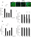
Figure 1. SuPAR down-modulates nephrin expression in human primary podocytes.
(a) Haematoxylin-eosin-staining (left picture) of human kidney from patients underwent to nephrectomy due to the renal cell carcinoma. Isolated human glomerula were cultured in vitro in order to obtain human primary podocytes (right picture). One representative experiment out of 20 is shown. (b) Quantification (left panel) of the immunoflourescence staining of nephrin expression in control (Mock) and human primary isolated podocytes treated with human recombinant suPAR (20 ng/mL) for 24 hours (suPAR). Results are expressed as MFI/cell and represent the average of 3 experiments ±SD. DAPI staining was used to determine nuclei number. Right picture represent one representative experiment out of 3 of nephrin expression in green (488 Alexa Fluor) in Mock and suPAR treated human primary podocytes. A nucleus staining is show in blue (DAPI). (c) Immunoflourescence staining of frozen tissue from human normal kidney with the specific clonal rabbit anti-nephrin Ab (488 Alexa Fluor). (d) QPCR analysis of nephrin expression by using specific human TaqMan assay in Mock and suPAR treated human podocytes. Results are expressed as relative fold change in suPAR treated cells vs Mock cells (ΔΔCt) and represent the average of 3 experiments ±SD. Values were normalized to GAPDH gene expression. Statistical significance (P) is indicated by asterisks and is represented as: **<0.01; ***<0.001.
Figure 2
Figure 2. Down-modulation of nephrin expression both at protein and transcription level in CIHPs.
(a) Quantification (left panel) of immunoflourescence staining (right upper panel) of nephrin expression in control (Mock) and CIHPs treated with different human recombinant suPAR for 24 hours (suPAR). Results are expressed as MFI/cell and represent the average of 6 experiments ±SD. DAPI staining was used to determine nuclei number. Results are expressed as MFI/cell and represent the average of 4 experiments ±SD. Right picture shows one representative immunoflourescence staining out of 4 of nephrin expression (488 Alexa Fluor) in green and nucleus (DAPI) in blue. (b) Dose-dependent qPCR analysis of nephrin expression in Mock and suPAR treated human podocytes by using specific human TaqMan assays. Results are expressed as relative fold change in suPAR treated cells vs Mock cells (ΔΔCt) and represent the average of 6 experiments ±SD. Values were normalized to the expression of GAPDH gene. (c) Time course qPCR analysis of nephrin and synaptopodin expression in Mock and suPAR treated human podocytes by using specific human TaqMan assays. Results are expressed as relative fold change in suPAR treated cells vs Mock cells (ΔΔCt) and represent the average of 6 experiments ±SD. Values were normalized to the expression of GAPDH gene. Statistical significance (P) is indicated by asterisks and is represented as: ***<0.001.
Figure 3
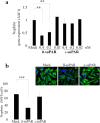
Figure 3. Full length DIDIIDIII suPAR down-regulates nephrin expression in CIHPs.
(a) QPCR analysis using specific human nephrin TaqMan assay in Mock and treated human podocytes with different concentration of full lengh suPAR (fl-suPAR) or short suPAR (c-suPAR) variants of suPAR. Results are expressed as relative fold change in suPAR treated cells vs Mock cells (ΔΔCt) and normalized to the expression of GAPDH gene. Results are represent as the average of 3 independent experiments ±SD. (b) Quantification of immunoflourescence staining (left panel) of nephrin expression in control (Mock) and treated human podocytes for 24 hours with 0.4 nM of the full-length (fl-suPAR) or cleaved (c-suPAR) variants of suPAR. Results are expressed as MFI/cell and represent the average of 3 experiments ±SD. Right picture shows one representative immunoflourescence staining out of 3 of nephrin expression (488 Alexa Fluor) in green and nucleus (DAPI) in blue.Statistical significance (P) is indicated by asterisks and is represented as: **<0.01; ***<0.001.
Figure 4
Figure 4. SuPAR mediated down-regulation of nephrin depends on αvβ3 integrin interaction and is associated with reduced activity of WT-1.
(a–b) Quantification of qPCR analysis of nephrin (a) and WT-1 (b) expression in Mock and suPAR treated (20 ng/mL) for 24 hours in CIHPs pre-incubated with different concentration (1, 5 and 10 μM) of αvβ3 antagonist (RGDfv). Results obtained by using the specify human TaqMan assays are expressed as relative fold change in treated cells vs. mock cells (ΔΔCt) and represent the average of 3 independent experiments ±SD. Values were normalized to the expression of GAPDH gene. Right upper picture of panel A shows one representative immunoflourescence staining out of 3 of αvβ3 (594 Alexa Fluor) integrin expression in red and nucleus in blue (DAPI) in CIHPs.
Figure 5
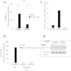
Figure 5. SuPAR induces lower WT-1 binding to the Nephrin gene promoter.
(a) Chromatin immunoprecipitation (ChIP) analysis of WT-1 binding to the cis region of Nphs1 gene promoter in CIHPs. Results are represented as fold change in the enrichment of precipitated chromatin fragments of Nphs1 gene promoter with either anti-WT-1 Ab or Rabbit Normal IgG in non treated (Mock) or treated with suPAR cells. Precipitated products were amplified by qPCR by using SYBR Green assay and normalized to DNA lacking any WT-1 site, located in the promoter of GAPDH gene of Input. (b) Binding of the specific anti-RNA polymerase II (CTD4H8) Ab to the DNA fragment of GAPDH gene was used as the positive control (Ctrl). Amplification of GAPDH gene promoter in the IgG and WT-1 chromatin immunoprecipitated of Mock and su-PAR treated samples were used as a negative Ctrl. ChIP samples were analyzed in triplicate and represented the average of 3 independent experiments ±SD. (c) QPCR analysis of WT-1 gene expression in Jurkat and K562 and CIHPs cell lines. TaqMan assays of 3 independent experiments ±SD. Values were normalized to the expression of GAPDH gene. (d) Detection of WT-1 protein by Western blotting assay (WB) after the chromatin immunoprecipitation with IgG control or anti-WT-1 antibodies in Jurkat and K562 cells. Analysis of β-actin of Inputs were used for normalization. One representative cropped blot out of two is shown. Statistical significance (P) is indicated by asterisks and is represented as: *<0.05; **<0.01; ***<0.001.
Figure 6
Figure 6. Injection of high doses of recombinant mouse suPAR into uPAR-knockout (Plaur−/−) mouse model induces down-regulation of nephrin expression.
(a) Quantification (left panel) of the ratio between urine total protein (mg)/creatinine (mg) concentration of suPAR treated mice with high dose of 20 μg of mouse recombinant for 24 hours vs control mice (Mock) (N = 3 mice for group). Immune-fluorescence in green (right panel) of suPAR (488 Alexa Fluor) deposit into glomerular tissue of suPAR treated Plaur−/− mice. (b) Quantification (left panel) of immunoflourescence staining of nephrin and synaptopodin expression in Mock and suPAR treated mice. (N = 3 mice for group). DAPI staining was used to determine cell numbers. Data are expressed as average of MFI/cell ±SD. Representative immunoflourescence staining (right panel) of nephrin in green (488 Alexa Fluor), synaptopodin in red (594 Alexa Fluor) and nucleus in blue (DAPI) expression in untreated (Mock) and suPAR treated mice (N = 3 mice for group). (c) QPCR analysis of nephrin and WT-1 expression in Mock and suPAR treated mice obtained by using specific mice TaqMan assays and expressed as relative fold change ±SD vs. mock cells. (N = 3 mice for group). Statistical significance (P) is indicated by asterisks and is represented as: *<0.05; **<0.01; ***<0.001.

References

    1. Sidenius N., Sier C. F. & Blasi F. Shedding and cleavage of the urokinase receptor (uPAR): identification and characterisation of uPAR fragments in vitro and in vivo. FEBS Lett 475, 52–56 (2000). - PubMed
    1. Thuno M., Macho B. & Eugen-Olsen J. suPAR: the molecular crystal ball. Dis Markers 27, 157–172 (2009). - PMC - PubMed
    1. Eden G., Archinti M., Furlan F., Murphy R. & Degryse B. The urokinase receptor interactome. Curr Pharm Des 17, 1874–1889 (2011). - PubMed
    1. Selleri C. et al. Involvement of the urokinase-type plasminogen activator receptor in hematopoietic stem cell mobilization. Blood 105, 2198–2205 (2005). - PubMed
    1. Montuori N. & Ragno P. Multiple activities of a multifaceted receptor: roles of cleaved and soluble uPAR. Front Biosci (Landmark Ed) 14, 2494–2503 (2009). - PubMed

Publication types

MeSH terms