Randomized Controlled Trial
. 2016 Jul;86(4):590-8.
doi: 10.2319/051115-319.1. Epub 2015 Sep 18.

Effects of different rapid maxillary expansion appliances on facial soft tissues using three-dimensional imaging

Affiliations
Randomized Controlled Trial

Effects of different rapid maxillary expansion appliances on facial soft tissues using three-dimensional imaging

Sedat Altındiş et al. Angle Orthod. 2016 Jul.

Abstract

Objective: To determine three-dimensional (3D) effects of three different rapid maxillary expansion (RME) appliances on facial soft tissues.

Materials and methods: Forty-two children (18 boys, 24 girls) who required RME treatment were included in this study. Patients were randomly divided into three equal groups: banded RME, acrylic splint RME, and modified acrylic splint RME. For each patient, 3D images were obtained before treatment (T1) and at the end of the 3-month retention (T2) with the 3dMD system.

Results: When three RME appliances were compared in terms of the effects on the facial soft tissues, there were no significant differences among them. The mouth and nasal width showed a significant increase in all groups. Although the effect of the acrylic splint RME appliances on total face height was less than that of the banded RME, there was no significant difference between the appliances. The effect of the modified acrylic splint appliance on the upper lip was significant according to the volumetric measurements (P < .01).

Conclusions: There were no significant differences among three RME appliances on the facial soft tissues. The modified acrylic splint RME produced a more protrusive effect on the upper lip.

Keywords: 3dMD; Face analysis; Rapid maxillary expansion; Soft tissue; Three-dimensional imaging.


Figures

Figure 1. Appliances used in this study. (a) Banded. (b) Acrylic cap splint bonded. (c) Modified acrylic cap splint bonded.
Figure 1.
Appliances used in this study. (a) Banded. (b) Acrylic cap splint bonded. (c) Modified acrylic cap splint bonded.
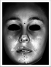
Figure 2. Anthropometric landmarks of soft tissue. n, indicates nasion; prn, pronasale; sn, subnasale; all-alr, left and right alare; c, columella; ls, labiale superius; stos, stomion superius; stoi, stomion inferius; li, labiale inferius; chr-chl, left and right cheilion; sl, sublabiale; pog, pogonion; gn, gnathion.
Figure 2.
Anthropometric landmarks of soft tissue. n, indicates nasion; prn, pronasale; sn, subnasale; all-alr, left and right alare; c, columella; ls, labiale superius; stos, stomion superius; stoi, stomion inferius; li, labiale inferius; chr-chl, left and right cheilion; sl, sublabiale; pog, pogonion; gn, gnathion.
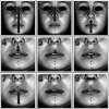
Figure 3. Linear measurements of soft tissue. (1) Total face height (n-gn). (2) Upper face height (n-stos). (3) Lower face height (sn-gn). (4) Upper lip height (sn-stos). (5) Upper vermillion height (ls-stos). (6) Lower vermillion height (stoi-li). (7) Lower lip height (stoi-gn). (8) Mouth width (chr-chl). (9) Nasal width (alr-all).
Figure 3.
Linear measurements of soft tissue. (1) Total face height (n-gn). (2) Upper face height (n-stos). (3) Lower face height (sn-gn). (4) Upper lip height (sn-stos). (5) Upper vermillion height (ls-stos). (6) Lower vermillion height (stoi-li). (7) Lower lip height (stoi-gn). (8) Mouth width (chr-chl). (9) Nasal width (alr-all).
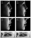
Figure 4. Angular measurements of soft tissue. (1) Total convexity angle (n-prn-pog). (2) Convexity angle (n-sn-pog). (3) Nasolabial angle (c-sn-ls). (4) Labiomental angle (li-sl-pog). (5) Upper lip angle (chr-ls-chl). (6) Lower lip angle (chr-li-chl).
Figure 4.
Angular measurements of soft tissue. (1) Total convexity angle (n-prn-pog). (2) Convexity angle (n-sn-pog). (3) Nasolabial angle (c-sn-ls). (4) Labiomental angle (li-sl-pog). (5) Upper lip angle (chr-ls-chl). (6) Lower lip angle (chr-li-chl).
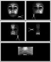
Figure 5. Determination of region of interest area for upper lip volumetric calculation.
Figure 5.
Determination of region of interest area for upper lip volumetric calculation.

References

    1. Haas AJ. The treatment of maxillary deficiency by opening the midpalatal suture. Angle Orthod. 1965;35:200–217. - PubMed
    1. Wertz RA. Skeletal and dental changes accompanying rapid midpalatal suture opening. Am J Orthod. 1970;58:41–66. - PubMed
    1. Zimring JF, Isaacson RJ. Forces produced by rapid maxillary expansion: III. Forces present during retention. Angle Orthod. 1965;35:178–186. - PubMed
    1. Kanomi R, Deguchi T, Kakuno E, Takano-Yamamoto T, Roberts WE. CBCT of skeletal changes following rapid maxillary expansion to increase arch-length with a development-dependent bonded or banded appliance. Angle Orthod. 2013;83:851–857. - PMC - PubMed
    1. Braun S, Bottrel A, Lee KG, Lunazzi JJ, Legan H. The biomechanics of rapid maxillary sutural expansion. Am J Orthod Dentofacial Orthop. 2000;118:257–261. - PubMed

Publication types

LinkOut - more resources