Review
. 2016 May;1863(5):892-901.
doi: 10.1016/j.bbamcr.2015.09.014. Epub 2015 Sep 14.

De novo peroxisome biogenesis: Evolving concepts and conundrums

Affiliations
Review

De novo peroxisome biogenesis: Evolving concepts and conundrums

Gaurav Agrawal et al. Biochim Biophys Acta. 2016 May.

Abstract

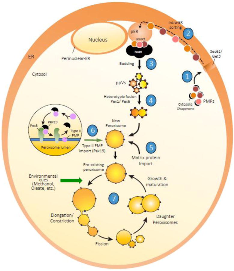
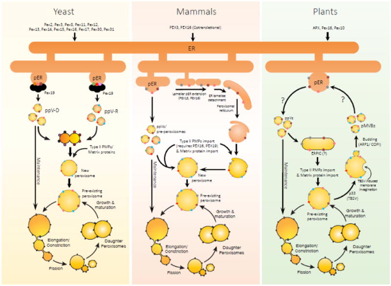
Peroxisomes proliferate by growth and division of pre-existing peroxisomes or could arise de novo. Though the de novo pathway of peroxisome biogenesis is a more recent discovery, several studies have highlighted key mechanistic details of the pathway. The endoplasmic reticulum (ER) is the primary source of lipids and proteins for the newly-formed peroxisomes. More recently, an intricate sorting process functioning at the ER has been proposed, that segregates specific PMPs first to peroxisome-specific ER domains (pER) and then assembles PMPs selectively into distinct pre-peroxisomal vesicles (ppVs) that later fuse to form import-competent peroxisomes. In addition, plausible roles of the three key peroxins Pex3, Pex16 and Pex19, which are also central to the growth and division pathway, have been suggested in the de novo process. In this review, we discuss key developments and highlight the unexplored avenues in de novo peroxisome biogenesis.

Keywords: De novo peroxisome biogenesis; Intra-ER sorting, import of PMPs into the ER, growth and division vs de novo peroxisome biogenesis; Pre-peroxisomal vesicles, ppVs; Pre-peroxisomal-ER, pER; Role of endoplasmic reticulum in peroxisome biogenesis, peroxisomal membrane protein biogenesis; Role of the endoplasmic reticulum in yeast, mammals and plants.


Figures

Figure 1
Figure 1
Schematic representation of peroxisome biogenesis pathways 1[ PMPs are both translated in the cytosol on free ribosomes or on the ER-associated ribosomes and incorporated post-translationally or cotranslationally in the ER-membrane. The ER-translocon, Sec61, is important for the PMP incorporation process. Similarly, TA-proteins are imported into the ER-membrane via the GET pathway. 2[ Subsequently, an intra-ER sorting process targets the PMPs to respective pER domains. 3[ The PMPs are exported from the ER in vesicular carriers and require Pex19. Pex16 is also important for the exit of Pex3 and other PMPs from the ER in mammalian cells. 4[ The vesicular carriers containing complementary sets of PMPs fuse to assemble the importomer complex. The fusion process requires peroxins Pex1 and Pex6. 5[ This assembly enables the nascent peroxisome to import matrix proteins and become a metabolically active organelle. 6[ Type II PMPs are imported directly into the peroxisome membrane with the assistance of Pex3 and Pex19 (Inset). 7[ The e novo route involving the ER also contributes to the cellular peroxisome population, thus sustaining the growth and division pathway and substituting for it when it is blocked or impaired. Using this backup pathway, in mutant cells (such as pex3Δ and pex19Δ cells) lacking functional pre-existing peroxisomes, reintroduction of the missing gene will form peroxisomes e novo and the new peroxisomes that are generated will restart growth and division pathway.
Figure 2
Figure 2
Comparative account of the ER-derived route of peroxisome biogenesis in yeast, mammalian cells and plants: The contribution of the ER to peroxisome biogenesis is evident in different model organisms, however, there are distinct mechanistic aspects specific for each system. In yeast, more PMPs have been documented to enter the ER than in any other system (section 3.1). As noted in Figure 1, these PMPs are sorted to the pER compartment. Recent results show that components of the peroxisomal RING and docking subcomplex proteins are sorted into distinct carriers (ppVs) [35]. In mammalian cells, such ER-derived ppVs are not yet described, however, pre-peroxisomal structures have been identified that sequentially import membrane proteins when peroxisome biogenesis is restored upon introduction of the missing protein in mutant cells [70] (section 3.2). In addition, these pre-peroxisomal structures presumably fuse with the pre-existing peroxisomes to replenish the growing and dividing peroxisomes. Various morphological studies depicted a close association of peroxisomes with specialized smooth-ER extensions also known as lamellar-ER [104,105]. This is distinct from the pER structures, which are associated with rough-ER. Pex13 is sorted to these lamellar-ER extensions and later detaches from the ER to form tubular structures. Once formed, additional integral membrane proteins are recruited leading to the assembly of the peroxisomal protein import apparatus. The formation of functional peroxisomes is concluded with the import of matrix enzymes and other Type II PMPs from the cytosol. Plants are the least studied system and trafficking of just a handful set of PMPs, including APX, Pex16 and Pex10, via the ER has been examined (section 3.3). However, neither ER-import requirements nor any peroxisome-specific vesicular carriers have been precisely defined in plants. None of the plant peroxin mutants have resulted in the complete loss of peroxisomes, thus the only proven pathway for peroxisome biogenesis is the growth and division model. A few morphological studies have noted that a non-ER, non-Golgi and non-peroxisomal compartment in plant cells receives Pex16 in cells recovering from BFA. This compartment is termed as ERPIC (ER-peroxisomes intermediate compartment), which in most ways act as a pre-peroxisomal compartment and is expected to be an intermediate in the formation of downstream peroxisomes. However, no concrete evidence exists delineating the functional significance of this compartment. In addition, during unique peroxisome to ER retrograde trafficking pathway has been identified in plants. Upon TBSV infection, plant peroxisomes undergo inward vesiculation resulting in the formation of pMBVs that traffics PMPs back to the pER. The mechanism by which these vesicle traffic PMPs back to the ER is unclear.

References

    1. Rhodin J. Correlation of Ultrastructural Organization and Function in Normal and Experimentally Changed Proximal Convoluted Tubule Cells of the Mouse Kidney: An Electron Microscopic Study. Including an Experimental Analysis of the Conditions for Fixation of the Renal Tissue for High Resolution Electron Microscopy. 1954;3:187–206.
    1. Bernhard WW, Rouiller CC. Microbodies and the problem of mitochondrial regeneration in liver cells. J Biophys Biochem Cytol. 1956;2:355–360. - PMC - PubMed
    1. De Duve C. Intracellular localization of enzymes. Nature. 1960;187:836–853. doi: 10.1038/187836a0. - DOI
    1. De Duve C. Functions of microbodies (peroxisomes) J Cell Biol. 1965;27:25A–26A. - PubMed
    1. Subramani S. Components involved in peroxisome import, biogenesis, proliferation, turnover, and movement. Physiol Rev. 1998;78:171–188. - PubMed

Publication types

MeSH terms

LinkOut - more resources