Review
. 2015 Oct;5(10):1024-39.
doi: 10.1158/2159-8290.CD-15-0507. Epub 2015 Sep 17.

MYC, Metabolism, and Cancer

Affiliations
Review

MYC, Metabolism, and Cancer

Zachary E Stine et al. Cancer Discov. 2015 Oct.

Abstract

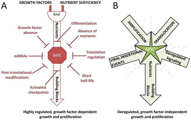
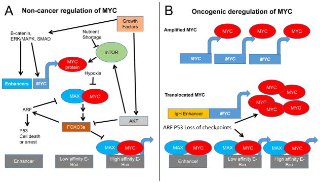
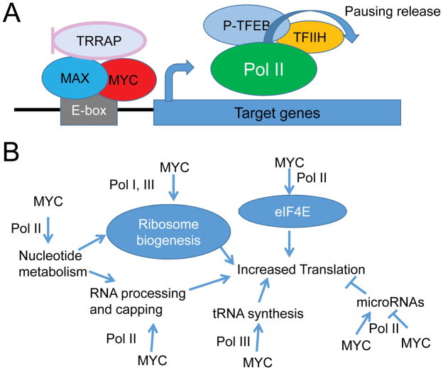
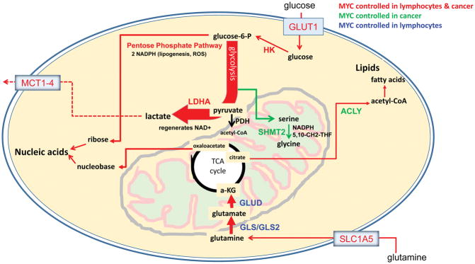
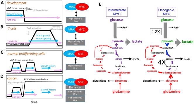
The MYC oncogene encodes a transcription factor, MYC, whose broad effects make its precise oncogenic role enigmatically elusive. The evidence to date suggests that MYC triggers selective gene expression amplification to promote cell growth and proliferation. Through its targets, MYC coordinates nutrient acquisition to produce ATP and key cellular building blocks that increase cell mass and trigger DNA replication and cell division. In cancer, genetic and epigenetic derangements silence checkpoints and unleash MYC's cell growth- and proliferation-promoting metabolic activities. Unbridled growth in response to deregulated MYC expression creates dependence on MYC-driven metabolic pathways, such that reliance on specific metabolic enzymes provides novel targets for cancer therapy.

Significance: MYC's expression and activity are tightly regulated in normal cells by multiple mechanisms, including a dependence upon growth factor stimulation and replete nutrient status. In cancer, genetic deregulation of MYC expression and loss of checkpoint components, such as TP53, permit MYC to drive malignant transformation. However, because of the reliance of MYC-driven cancers on specific metabolic pathways, synthetic lethal interactions between MYC overexpression and specific enzyme inhibitors provide novel cancer therapeutic opportunities.


Conflict of interest statement

The authors declare no conflict of interest.

Conflicts of Interest

No potential conflicts of interest were disclosed.

Figures

Figure 1
Figure 1. Schematic illustration of growth factor-dependent and growth factor-independent MYC activity
A) In non-cancerous cells, growth signals and adequate nutrients are required for MYC activity. Multiple levels of feedback loops and checkpoints further control MYC activity. B) In cancerous cells, in contrast, checkpoint loss, gene amplification, chromosomal translocation, abnormal enhancer activation, or one or more other deregulated signaling events lead to growth factor-independent MYC metabolic activities and subsequent unconstrained cellular growth and proliferation.
Figure 2
Figure 2
MYC regulation in non-cancerous and cancerous cells. A) In non-cancerous cells MYC expression is activated by growth factors through activation of enhancers. MYC protein, whose translation is enhanced by activated mTOR, dimerizes with MAX to form a heterodimer that activates transcription of genes containing high affinity E-boxes. Upon nutrient shortage or hypoxia, MYC translation, protein stability and MYC/MAX dimerization inhibited. Over-activation of MYC activates the ARF and p53 checkpoints resulting in cell death or arrest, while ARF can inhibit MYC function. Downstream of AKT, FOXO3a proteins counteract MYC activation. B) In cancer cells, constitutive activation of growth factor and mTOR signaling, loss of checkpoints, engagement of atypical enhancers, or amplification or translocation of MYC can increase levels of MYC to supraphysiologic levels independently of growth factors, causing MYC/MAX binding to lower affinity binding sites and enhancers in addition to high affinity sites. Loss of ARF or p53 checkpoints allows uncontrolled cell growth.
Figure 3
Figure 3
MYC enhances transcription and translation. A) The MYC/MAX dimer binds to E-boxes or lower affinity degenerate sequences to recruit histone acetylases or promote polymerase phosphorylation, thus release polymerase from pausing to amplify transcription. B) Acting on Pol I, Pol II and Pol III, MYC controls translation through upregulation of transcription of ribosomal subunits, tRNA, and nucleotide synthesis genes. MYC also stimulates translation by upregulating eukaryotic translation initiation factor 4E (eIF4E) and stimulating enzymes that control RNA processing and capping. MYC upregulation and downregulation of microRNAs also regulates the translation of microRNA targets.
Figure 4
Figure 4
Myc-regulated metabolic pathways in cancer. Glucose is taken up by glucose transporters (GLUT) and phosphorylated by hexokinase (HK) to form glucose-6-phosphate (glucose-6-P). Glucose-6-P can then either enter glycolysis or the pentose phosphate pathway, which supports nucleotide synthesis by yielding two NADPH reducing equivalents and one ribose per molecule of glucose. The serine biosynthesis pathway branches off glycolysis, producing serine and glycine that likewise support nucleotide synthesis. Serine hydroxymethyltransferase 2 (SHMT2) converts serine to glycine which, in a series of coupled reactions, can be used to create nucleotide and epigenetic methyl donor 5,10-CH2-tetrahydrofolate and mitochondrial NADPH for redox control. Lactate dehydrogenase (LDHA) can regenerate NAD+ by converting glycolysis-derived pyruvate to lactate, which is then exported out of the cell by monocarboxylate transporters (MCT1-4). Alternatively, pyruvate can enter the TCA cycle in a pyruvate dehydrogenase (PDH)-dependent conversion to acetyl-CoA. TCA cycle citrate can be exported to the cytoplasm where it is converted to acetyl-CoA by ATP citrate lyase (ACLY). Cytoplasmic acetyl-CoA can then be channeled into lipogenesis. In addition to glucose, glutamine is an important fuel source in cancers. Glutamine is transported across the membrane by the glutamine transporter (SLC1A5/ASCT2) and converted to glutamate by glutaminase (GLS or GLS2). Glutamate can then be converted to the TCA cycle intermediate α-ketoglutarate (αKG) by glutamate dehydrogenase (GLUD) or aminotransferases.
Figure 5
Figure 5
The effect of nutrient and growth factor availability on MYC driven metabolism. A) During cellular differentiation, loss of growth factor stimulation can turn off MYC- driven metabolism even in the presence of nutrients. B) In T cells, receptor stimulation and growth factors can drive MYC signaling, but withdraw of receptor stimulation and checkpoints turn off MYC-driven metabolism even in the presence of nutrients. C) In normal proliferating cells, MYC-driven metabolism is activated in the presence of both nutrients and growth factors, but individual exposure to nutrients or growth factors is not sufficient to activate MYC-driven metabolism. D) In cancer, MYC deregulation and loss of checkpoints leaves cells unable to turn off MYC-driven metabolism independent of growth factors and nutrient availability. The inability to turn off MYC-driven metabolism creates therapeutic vulnerabilities to metabolic inhibitors. E) Comparison of glucose and glutamine metabolism of cells expressing intermediate levels of MYC and oncogenic levels of MYC shows oncogenic levels of MYC cause small increase (1.2 fold) in lactate production and a large increase (4 fold) in TCA cycle flux (197).

Similar articles

Cited by

References

    1. Conacci-Sorrell M, McFerrin L, Eisenman RN. An overview of MYC and its interactome. Cold Spring Harbor perspectives in medicine. 2014;4:a014357. - PMC - PubMed
    1. Duesberg PH, Bister K, Vogt PK. The RNA of avian acute leukemia virus MC29. Proceedings of the National Academy of Sciences of the United States of America. 1977;74:4320–4. - PMC - PubMed
    1. Sheiness D, Fanshier L, Bishop JM. Identification of nucleotide sequences which may encode the oncogenic capacity of avian retrovirus MC29. Journal of virology. 1978;28:600–10. - PMC - PubMed
    1. Bister K, Jansen HW. Oncogenes in retroviruses and cells: biochemistry and molecular genetics. Advances in cancer research. 1986;47:99–188. - PubMed
    1. Dalla-Favera R, Bregni M, Erikson J, Patterson D, Gallo RC, Croce CM. Human c-myc onc gene is located on the region of chromosome 8 that is translocated in Burkitt lymphoma cells. Proceedings of the National Academy of Sciences of the United States of America. 1982;79:7824–7. - PMC - PubMed

Publication types

MeSH terms

Substances