. 2015 Sep 18;10(9):e0138127.
doi: 10.1371/journal.pone.0138127. eCollection 2015.

Green-to-Red Photoconversion of GCaMP

Affiliations

Green-to-Red Photoconversion of GCaMP

Minrong Ai et al. PLoS One. .

Abstract

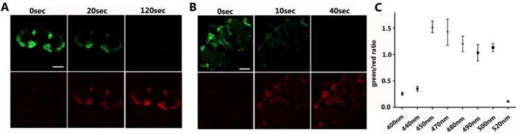
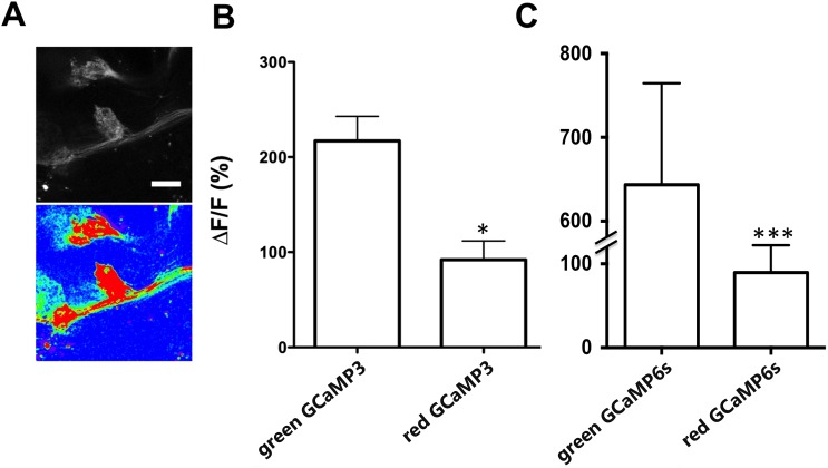
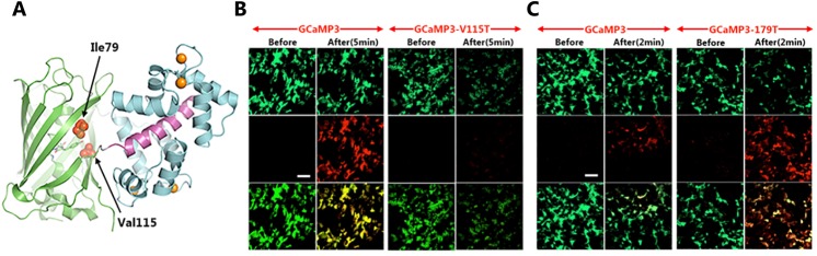
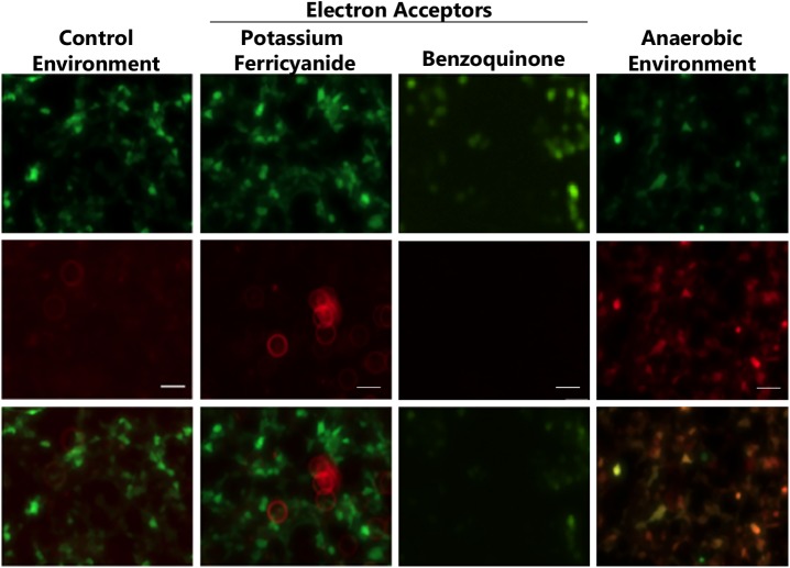
Genetically encoded calcium indicators (GECIs) permit imaging intracellular calcium transients. Among GECIs, the GFP-based GCaMPs are the most widely used because of their high sensitivity and rapid response to changes in intracellular calcium concentrations. Here we report that the fluorescence of GCaMPs--including GCaMP3, GCaMP5 and GCaMP6--can be converted from green to red following exposure to blue-green light (450-500 nm). This photoconversion occurs in both insect and mammalian cells and is enhanced in a low oxygen environment. The red fluorescent GCaMPs retained calcium responsiveness, albeit with reduced sensitivity. We identified several amino acid residues in GCaMP important for photoconversion and generated a GCaMP variant with increased photoconversion efficiency in cell culture. This light-induced spectral shift allows the ready labeling of specific, targeted sets of GCaMP-expressing cells for functional imaging in the red channel. Together, these findings indicate the potential for greater utility of existing GCaMP reagents, including transgenic animals.


Conflict of interest statement

Competing Interests: A patent application for this work has been filed, Green-to-red photo-convertible fluorescent calcium indicator, application number 14/622,025. There are no further patents, products in development or marketed products to declare. This does not alter the authors' adherence to all the PLOS ONE policies on sharing data and materials, as detailed online in the guide for authors.

Figures

Fig 1
Fig 1. GCaMP3 is converted from green to red fluorescence after exposure to blue light.
(A-B) Fluorescent micrographs of dissected Drosophila brains expressing UAS-GCaMP3; IR8a-GAL4 (A) or UAS-GCaMP6s; NP225-GAL4 (B). The dissected brains were exposed to blue light (mercury arc light passed through a Zeiss 63x oil-immersion objective) for different durations as indicated at the top of each panel. Green (top) and red (bottom) fluorescence micrographs were captured using a confocal microscope. Scale bar: 20μm. (C) Green-to-red photoconversion of GCaMP3 as quantified by the ratio of red-to-green fluorescence intensity (y-axis) following exposure to light from a Xenon lamp (x-axis).
Fig 2
Fig 2. Photoconverted GCaMP3 and GCaMP6s retain calcium sensitivity.
Calcium imaging of a dissected fly brain expressing IR8a-GAL4 and UAS-GCaMP3 in response to depolarizing reagent, 40mM KCl. Images of red fluorescence from a brain before (top panel) and after (bottom panel, pseudo-colored) KCl stimulation are shown. Scale bar: 10μm. (B) Quantification of changes in GCaMP3 fluorescent intensity (ΔF/F) in response to KCl depolarization. (C) Fluorescent intensity (ΔF/F) in response to KCl was measured in the brains of flies harboring UAS-GCaMP6s and NP225-GAL4, which expresses in central neurons. Student’s t-test. Error bars represent SEM. *p<0.05; ***p<0.01. n > 4 for each experimental group.
Fig 3
Fig 3. The amino acid residues critical for green-to-red photoconversion.
(A) A 3-D crystal structure of GCaMP3 protein illustrating GFP backbone in green, calmodulin in cyan and myosin M13 in purple. The spatial positions of amino acid Ile79 and Val115 are highlighted by arrows. (B-C) Control and mutant GCaMP3s were expressed in HEK293 cells. Green (top), red (middle) and merged (bottom) fluorescent micrographs were taken before and after the cells were exposed to blue light (mercury arc light passed through a FITC filter) for either 5 minutes (B) or 2 minutes (C). Note that V115T results in a loss of green-to-red photoconversion, whereas I79T improves efficiency. Scale bar: 20μm.
Fig 4
Fig 4. The efficiency of GCaMP3 photoconversion in HEK293 cells is dramatically enhanced under low-oxygen condition.
GCaMP3-expressing HEK293 cells were illuminated first under control culture conditions, with the presence of electron acceptor potassium ferricyanide or benzoquinone (both at 5mM), or under low-oxygen conditions created by treating the cells with 30μg/ml catalase, 4.5mg/ml glucose and 250μg/ml glucose oxidase for 30 minutes. Cells were then exposed to blue light for 2 minutes. Green (top), red (middle) and merged (bottom) fluorescent micrographs. Scale bar: 20μm.

References

    1. Miyawaki A, Llopis J, Heim R, McCaffery JM, Adams JA, Ikura M, et al. Fluorescent indicators for Ca2+ based on green fluorescent proteins and calmodulin. Nature. 1997;388(6645):882–7. 10.1038/42264 . - DOI - PubMed
    1. Nakai J, Ohkura M, Imoto K. A high signal-to-noise Ca(2+) probe composed of a single green fluorescent protein. Nat Biotechnol. 2001;19(2):137–41. 10.1038/84397 . - DOI - PubMed
    1. Patterson GH, Lippincott-Schwartz J. A photoactivatable GFP for selective photolabeling of proteins and cells. Science. 2002;297(5588):1873–7. 10.1126/science.1074952 . - DOI - PubMed
    1. Ando R, Hama H, Yamamoto-Hino M, Mizuno H, Miyawaki A. An optical marker based on the UV-induced green-to-red photoconversion of a fluorescent protein. Proc Natl Acad Sci U S A. 2002;99(20):12651–6. 10.1073/pnas.202320599 - DOI - PMC - PubMed
    1. Wiedenmann J, Ivanchenko S, Oswald F, Schmitt F, Rocker C, Salih A, et al. EosFP, a fluorescent marker protein with UV-inducible green-to-red fluorescence conversion. Proc Natl Acad Sci U S A. 2004;101(45):15905–10. 10.1073/pnas.0403668101 - DOI - PMC - PubMed

Publication types

MeSH terms

LinkOut - more resources