Review
. 2016 Jan;22 Suppl 1(0 1):S29-33.
doi: 10.1016/j.parkreldis.2015.09.020. Epub 2015 Sep 8.

Tauopathies as clinicopathological entities

Affiliations
Review

Tauopathies as clinicopathological entities

David J Irwin. Parkinsonism Relat Disord. 2016 Jan.

Abstract

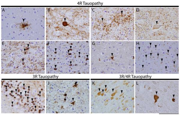
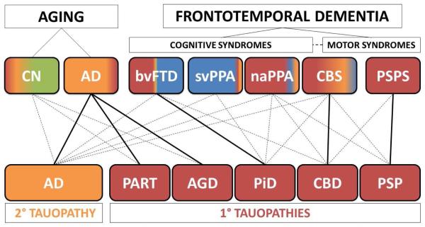
Tauopathies are a class of neurodegenerative disorders characterized by neuronal and/or glial inclusions composed of the microtubule-binding protein, tau. Several lines of evidence suggest tau aggregation is central to the neurodegenerative process in tauopathies. First, recent animal and cell model studies find abnormally-modified tau alone may be transmitted between adjacent neurons and spread to anatomically connected brain regions to recapitulate human disease. Further, staging efforts in human autopsy cases suggest a sequential distribution of tau aggregation in the central nervous system that could reflect this observed cell-to-cell transmission of pathogenic tau species in animal models. Finally, pathogenic mutations in the MAPT gene encoding tau protein cause hereditary forms of tauopathy. Clinically, tauopathies can present with a range of phenotypes that include both movement- and cognitive/behavioral-disorders (i.e. frontotemporal dementia spectrum disorders) or non-specific amnestic symptoms in advanced age. A major limitation is that current clinical diagnostic criteria for these disorders do not reliably differentiate underlying tauopathy from other neurodegenerative diseases, such as TDP-43 proteinopathies. Thus, current research efforts are focused on improving the ante mortem diagnosis of tauopathies, including pre-clinical stages of disease, as many therapeutic strategies for emerging disease-modifying therapies focus on preventing abnormal folding and spread of tau pathology.

Keywords: Argyrophilic grain disease; Corticobasal degeneration; Corticobasal syndrome tauopathy; Frontotemporal dementia; Frontotemporal lobar degeneration; MAPT mutation; Pick's disease; Primary age related tauopathy; Primary progressive aphasia; Progressive supranuclear palsy.


Figures

Figure 1
Figure 1. Tauopathy histopathological subtypes
Photomicrographs of illustrate characteristic tau inclusion morphologies for 4R (a-h), 3R (I,J) and 3R/4R (K,L) primary tauopathies. Characteristic PSP tauopathy including (A) a tufted astrocytes in mid-frontal cortex and (B) a globose neuronal tau tangle in midbrain. CBD characteristic tau pathology showing (C) glial astrocytic plaques in the mid-frontal cortex, (D) a ballooned neuron in the anterior cingulate gyrus and (E) oligodendrocyte coiled bodies in the mid-frontal cortex. Note ballooned neurons and coiled bodies may be present in other tauopathies to a lesser extent. GGT (F) globular oligodendrocytes in white matter and (G) astrocytic tau inclusions in grey matter of the mid-frontal cortex show distinct globular morphology from PSP and CBD tau pathology. AGD associated (H) small comma-shaped 4R tau reactive grains (arrows) in the amygdala. PiD (I) spherical “Pick bodies” seen in the hippocampal dentate gyrus neurons and (J) mid-frontal cortex (asterisks), along with glial tau inclusions (arrows). PART (K) neurofibrillary tau tangles seen in the cornu ammonis of the hippocampus. ALS-PDC spinal cord with (L) tau-reactive tangles in lower motor neurons of the spinal cord. PHF-1 stain. Scale bar=100 µm.
Figure 2
Figure 2. Clinicopathological correlations in tauopathies
The scheme portrays (top) the relative frequencies of neuropathological subtypes of FTLD-Tau (i.e. primary tauopathies-red) and AD (i.e. secondary tauopathy-yellow) seen at autopsy in clinical phenotypes in FTD-spectrum disorders and aging by the color-coding of each box. Unremarkable neuropathological findings are shaded in green and TDP-43 protienopathies in blue (this schematic does not account for cerebrovascular changes, co-morbid pathology or less common neurodegenerative diseases). FTD clinical phenotypes are divided by cognitive and motor syndromes; the dashed line represents the clinical overlap between FTD cognitive and motor syndromes and CBS is placed intermediate to these categories as this clinical syndrome has aspects of both cognitive and motor dysfunction. AD neuropathology (yellow) is found in approximately a third of older patients who are cognitively normal (CN) and is seen in the majority amnestic AD clinical phenotype. FTLD-Tau (red) is found in virtually all PSPS cases and the majority of naPPA. FTLD-Tau is also found in a significant proportion of CBS and relatively rare in svPPA. Roughly half of bvFTD cases harbor FTLD-Tau while a small percentage can have AD neuropathology (yellow). Solid lines represent the predominant clinicopathological association for each specific tauopathy (bottom) while dashed lines represent less common clinical manifestations of each tauopathy. AD neuropathology is most commonly associated with the amnestic AD clinical phenotype but may also present as bvFTD, PPA variants and CBS. PART and AGD are largely associated with late onset (>80 years) amnestic syndrome similar to clinical AD and less commonly in CN individuals. PiD is most commonly found in association with bvFTD but can present with PPA variants or CBS. CBD tauopathy is primarily associated with CBS but also can manifest as bvFTD, naPPA or PSPS, while PSP is predominantly associated with PSPS and less commonly associated with naPPA or CBS.

References

    1. Mackenzie IR, Neumann M, Bigio EH, Cairns NJ, Alafuzoff I, Kril J, et al. Nomenclature and nosology for neuropathologic subtypes of frontotemporal lobar degeneration: an update. Acta neuropathologica. 2010;119:1–4. - PMC - PubMed
    1. Rascovsky K, Hodges JR, Knopman D, Mendez MF, Kramer JH, Neuhaus J, et al. Sensitivity of revised diagnostic criteria for the behavioural variant of frontotemporal dementia. Brain : a journal of neurology. 2011;134:2456–77. - PMC - PubMed
    1. Gorno-Tempini ML, Hillis AE, Weintraub S, Kertesz A, Mendez M, Cappa SF, et al. Classification of primary progressive aphasia and its variants. Neurology. 2011;76:1006–14. - PMC - PubMed
    1. Litvan I, Agid Y, Calne D, Campbell G, Dubois B, Duvoisin RC, et al. Clinical research criteria for the diagnosis of progressive supranuclear palsy (Steele-Richardson-Olszewski syndrome): report of the NINDS-SPSP international workshop. Neurology. 1996;47:1–9. - PubMed
    1. Armstrong MJ, Litvan I, Lang AE, Bak TH, Bhatia KP, Borroni B, et al. Criteria for the diagnosis of corticobasal degeneration. Neurology. 2013;80:496–503. - PMC - PubMed

Publication types

MeSH terms