. 2015 Sep 18;10(9):e0138625.
doi: 10.1371/journal.pone.0138625. eCollection 2015.

mTOR Overactivation and Compromised Autophagy in the Pathogenesis of Pulmonary Fibrosis

Affiliations

mTOR Overactivation and Compromised Autophagy in the Pathogenesis of Pulmonary Fibrosis

Yao-Song Gui et al. PLoS One. .

Abstract

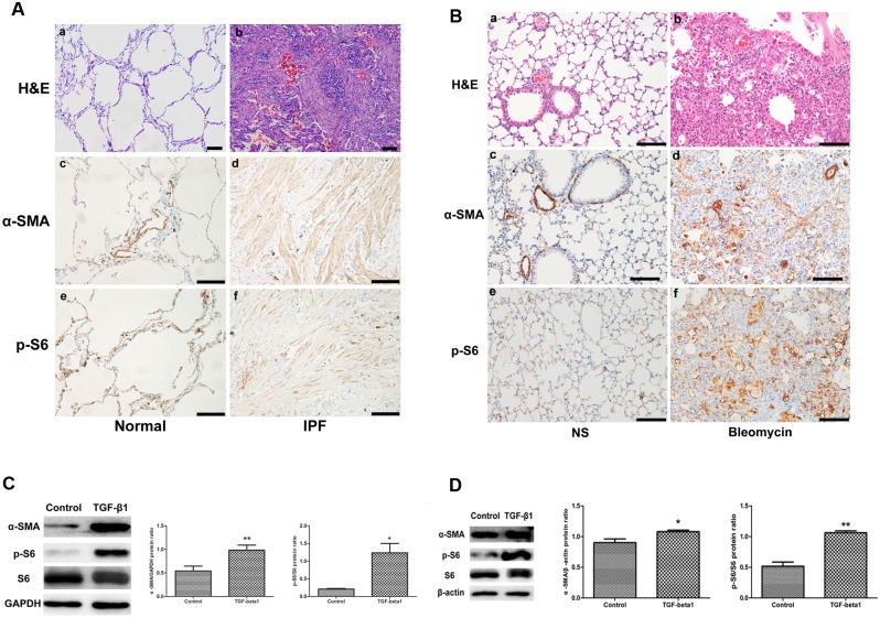
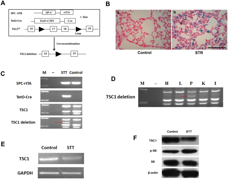
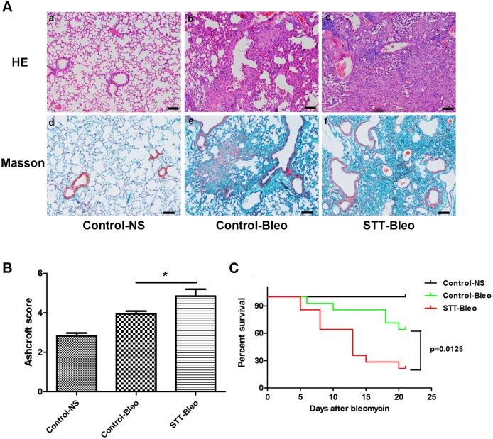
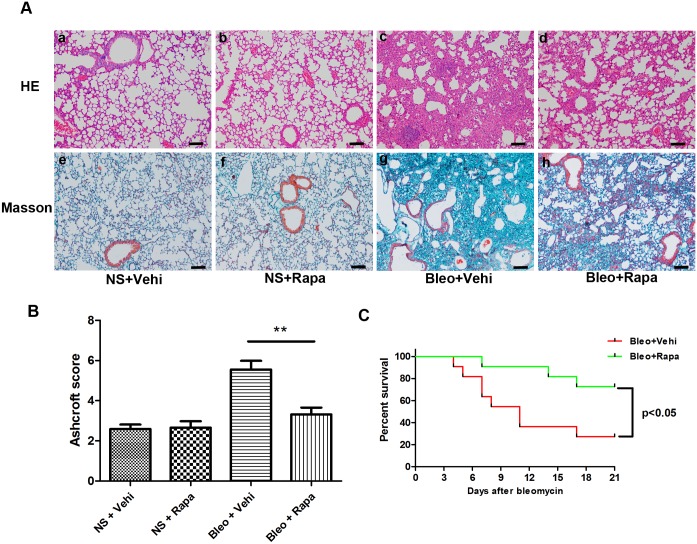
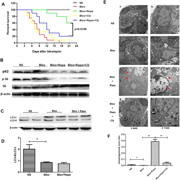
The mammalian target of rapamycin (mTOR) signaling pathway in pulmonary fibrosis was investigated in cell and animal models. mTOR overactivation in alveolar epithelial cells (AECs) was achieved in the conditional and inducible Tsc1 knock-down mice SPC-rtTA/TetO-Cre/Tsc1(fx/+) (STT). Doxycycline caused Tsc1 knock-down and consequently mTOR activation in AECs for the STT mice. Mice treated with bleomycin exhibited increased mortality and pulmonary fibrosis compared with control mice. In wild-type C57BL/6J mice, pretreatment with rapamycin attenuated the bleomycin-mediated mortality and fibrosis. Rapamycin-mediated mouse survival benefit was inhibited by chloroquine, an autophagy inhibitor. Autophagosomes were decreased in the lungs after bleomycin exposure. Rapamycin induced the production of autophagosomes and diminished p62. We concluded that mTOR overactivation in AECs and compromised autophagy in the lungs are involved in the pathogenesis of pulmonary fibrosis. The suppression of mTOR and enhancement of autophagy may be used for treatment of pulmonary fibrosis.


Conflict of interest statement

Competing Interests: The authors have declared that no competing interests exist.

Figures

Fig 1
Fig 1. mTOR was activated during the process of pulmonary fibrosis in vivo and vitro.
A) mTOR activation in fibroblast foci of lung tissue in IPF patients. a,b, H&E staining with normal control and IPF lung tissues; Immunohistochemical staining performed with α-SMA (c,d) and p-S6 (e,f) antibodies showed an increase in α-SMA and p-S6 in IPF lung tissues (d, f) compared with the control (c, e). Scale bar = 100 μm. B) mTOR activation in the lung tissues of C57BL/6J mice after bleomycin intra-tracheal injection. a,b, H&E staining with lungs of saline and bleomycin-treated mice; Immunohistochemical staining was performed with α-SMA (c,d) and p-S6 (e,f) antibodies in saline- and bleomycin-treated mouse lung tissues. NS, normal saline. Scale bar = 100 μm. C) mTOR signaling pathway was activated in primary lung fibroblasts isolated from normal controls treated with TGF-β1(5 ng/ml) for 48 h. Western blot analysis of α-SMA and p-S6 in control and TGF-β1-treated primary lung fibroblasts (a). Densitometric quantification of the Western blot in (a) is shown in (b) with α-SMA normalized against GAPDH and (c) with p-S6 normalized against S6. **, P<0.01; *, P<0.05. n = 3. D) mTOR signaling pathway was activated in MRC5 cells (a human fetal lung fibroblast cell line) treated with TGF-β1 (5 ng/ml) for 48 h. Western blot analysis of α-SMA and p-S6 in control and TGF-β1-treated MRC5 cells (a). Densitometric quantification of the Western blot in (a) is shown in (b) with α-SMA normalized against β-actin and (c) with p-S6 normalized against S6. **, p<0.01; *, p<0.05. n = 3.
Fig 2
Fig 2. Generation of inducible Tsc1 knock-down in lung alveolar epithelial cells (SPC-rtTA/TetO-Cre/Tsc1 f/+ (STT) mice).
A) Exons 17 and 18 of the Tsc1 gene were deleted in alveolar epithelial cells of 4-week-old STT mice by administering doxycycline (Dox). B) β-galactosidase activity was detected in lung alveolar epithelial cells from SPC-rtTA/TetO-Cre/ROSA26R (STR) transgenic mice after doxycycline treatment (b), while it could not be detected in control mice (a). Scale bar = 100 μm. C) DNA from lung tissues of STT transgenic mice treated with doxycycline were examined by PCR to detect Tsc1 knock-down, indicating Cre recombinase activity. Tsc1 deletion band (370 bp, red arrow) was detected in STT mice but not in control mice. M, DNA marker;-, water control. D) The Tsc1 deletion band (red arrow) was detected in the lungs but not in other organs of a STT transgenic mouse receiving doxycycline treatment. M, DNA marker;-, water control; H, heart; L, liver; P, pulmonary; K, kidney; I, intestine. E) Tsc1 gene knock-down was confirmed by RT-PCR for Tsc1 mRNA in lungs from a STT transgenic mouse after doxycycline treatment, and GAPDH was used as the control. F) TSC1 and p-S6 proteins were detected by Western blot in lungs of doxycycline-treated STT transgenic mice. TSC1 was decreased, and p-S6 was elevated, indicating TSC1 knock-down in the lungs of STT mice. S6 and β-actin were used as controls.
Fig 3
Fig 3. Conditional Tsc1 knock-down in lung alveolar epithelial cells from doxycyline-treated STT mice exacerbated bleomycin-mediated lung injury.
A) Histological analysis of lungs in the mice treated with bleomycin at day 21. H&E staining (a-c) and Masson’s trichrome staining (d-f) were performed. STT mice had more severe lung injury (c, f) than control mice (b, e). Scale bar = 100 μm. NS, saline; Bleo, bleomycin. B) Semi-quantitative assessment was performed on day 21 using Ashcroft scoring method, a significantly higher score was observed in STT mice treated with Bleo than control mice treated with Bleo. Results were expressed as mean±SEM, n = 6 mice per group, * p<0.05. C) STT mice had a higher mortality rate than control mice after a single intra-tracheal injection of bleomycin for 21 days.
Fig 4
Fig 4. Bleomycin-mediated lung injury in wild-type C57BL/6J mice was attenuated by rapamycin (treatment initiated at 5 days before bleomycin injection).
A) H&E staining (a-d) and Masson’s trichrome staining (e-h) of mouse lungs were performed after bleomycin injection at day 21. Lung injury was milder in rapamycin-treated mice (d, h) compared with vehicle-treated mice (c, g). Scale bar = 100 μm. NS, saline; Bleo, bleomycin; Vehi, vehicle; Rapa, rapamycin. B) Semi-quantitative assessment was performed on day 21 using Ashcroft scoring method, a significantly higher score was observed in the mice treated with Bleo (no rapamycin) than those treated with Bleo+Rapa. Results were expressed as mean±SEM, n = 6 mice per group, ** p<0.01. C) Bleomycin-mediated mouse mortality was decreased after rapamycin treatment.
Fig 5
Fig 5. Rapamycin-induced autophagyin the bleomycin-mediated lung injury and fibrosis model.
A) Rapamycin decreased the death caused by bleomycin. Chloroquine, an autophagy inhibitor, reversed the benefit of rapamycin in the bleomycin-mediated lung injury model (Bleo+Rapa+CQ vs Bleo+Rapa, p = 0.0158). B) Western blot analysis of p62 and p-S6 were performed in the bleomycin-mediated lung injury and fibrosis model. p62, a protein inversely correlated with autophagy activity, was decreased in lungs of mice treated with rapamycin alone. p62 expression was higher with combined rapamycin and chloroquine treatment than with rapamycin alone. S6 and β-actin were used as controls. C) Western blot ananlysis of LC3 I and LC3 II were performed in the bleomycin-mediated lung injury and fibrosis mice model. D) Relative density of LC3 II/LC3 I of bands in Fig 5C. Autophagy was significantly decreased in bleomycin-mediated lung injury and fibrosis model (*bleomycin vs normal saline, p < 0.05). E) Electron microscope images of lung tissues show autophagosomes in the bleomycin-mediated lung injury model. Arrows indicate autophagosomes. Rapamycin treatment alone induced an increased number of autophagosomes. Left panel, original magnification: 6,000X and right panel, original magnification: 11,500X. F) Statistical results for the autophagosomes in Fig 5E. The statistical results indicate the percent area of autophagosomes in a cell.

References

    1. Raghu G, Collard HR, Egan JJ, Martinez FJ, Behr J, Brown KK, et al. An official ATS/ERS/JRS/ALAT statement: idiopathic pulmonary fibrosis: evidence-based guidelines for diagnosis and management. American journal of respiratory and critical care medicine. 2011;183(6):788–824. Epub 2011/04/08. 10.1164/rccm.2009-040GL . - DOI - PMC - PubMed
    1. King TE Jr., Bradford WZ, Castro-Bernardini S, Fagan EA, Glaspole I, Glassberg MK, et al. A phase 3 trial of pirfenidone in patients with idiopathic pulmonary fibrosis. The New England journal of medicine. 2014;370(22):2083–92. Epub 2014/05/20. 10.1056/NEJMoa1402582 . - DOI - PubMed
    1. Richeldi L, du Bois RM, Raghu G, Azuma A, Brown KK, Costabel U, et al. Efficacy and safety of nintedanib in idiopathic pulmonary fibrosis. The New England journal of medicine. 2014;370(22):2071–82. Epub 2014/05/20. 10.1056/NEJMoa1402584 . - DOI - PubMed
    1. Dobashi Y, Watanabe Y, Miwa C, Suzuki S, Koyama S. Mammalian target of rapamycin: a central node of complex signaling cascades. International journal of clinical and experimental pathology. 2011;4(5):476–95. Epub 2011/07/09. - PMC - PubMed
    1. Hay N, Sonenberg N. Upstream and downstream of mTOR. Genes & development. 2004;18(16):1926–45. Epub 2004/08/18. 10.1101/gad.1212704 . - DOI - PubMed

Publication types