Review
. 2015 Oct;25(4):273-80.
doi: 10.1016/j.semradonc.2015.05.008. Epub 2015 May 14.

Patient-Derived Xenografts as a Model System for Radiation Research

Affiliations
Review

Patient-Derived Xenografts as a Model System for Radiation Research

Christopher D Willey et al. Semin Radiat Oncol. 2015 Oct.

Abstract

The cancer literature is filled with promising preclinical studies demonstrating impressive efficacy for new therapeutics, yet translation of these approaches into clinical successes has been rare, indicating that current methods used to predict efficacy are suboptimal. The most likely reason for the limitation of these studies is the disconnect between preclinical models and cancers treated in the clinic. Specifically, most preclinical models are poor representations of human disease. Immortalized cancer cell lines that dominate the cancer literature may be, in a sense, "paper tigers" that have been selected by decades of culture to be artificially driven by highly targetable proteins. Thus, although effective in treating these cell lines either in vitro or as artificial tumors transplanted from culture into experimental animals as xenografts, the identified therapies would likely underperform in a clinical setting. This inherent limitation applies not only to drug testing but also to experiments with radiation therapy. Indeed, traditional radiobiology methods rely on monolayer culture systems, with emphasis on colony formation and DNA damage assessment that may have limited clinical translation. As such, there has been keen interest in developing tumor explant systems in which patient tumors are directly transplanted into and solely maintained in vivo, using immunocompromised mice. These so-called patient-derived xenografts (PDXs) represent a robust model system that has been garnering support in academia and industry as a superior preclinical approach to drug testing. Likewise, PDX models have the potential to improve radiation research. In this review, we describe how PDX models are currently being used for both drug and radiation testing and how they can be incorporated into a translational research program.


Figures

Figure 1
Figure 1
Patient-Derived Xenograft Advantages and Challenges. Schematic of derivation of Patient-derived xenografts (PDX) is shown. Passage (P) number is indicated. The number of passages that can be utilized depends on the tumor's genetic stability as well as the host type. Athymic nude mice tend to maintain the PDX genetic stability longer than more severely immunocompromised mice though at the expense of lower take rates. At each passage, PDX tumor is typically frozen down. Advantages of PDX over immortalized cell lines are shown. Challenges that are encountered with a PDX program are listed.
Figure 2
Figure 2
Approaches to Radiation Biology Study using Patient-Derived Xenografts. Patient-derived xenograft (PDX) tumors and cells can potentially be used for radiation studies. PDX tumors can be directly tested in vivo for tumor growth and animal survival using mouse irradiators. With the advent of conformal irradiators such as the small animal radiation research platform (SARRP, Xstrahl and XRAD 225Cx, Precision X-ray), orthotopically implanted tumors can be specifically treated with image guidance and/or monitoring. PDX tumors are expected to contain cancer stem cells or stem cell-like cells (CSC's) that can be maintained as tumor spheres when cultured in non-differentiating media. An example of Glioblastoma multiforme (GBM) PDX neurospheres is shown. PDX tumor cells can also be implanted into extracellular matrix material as 3D culture. As with tumor spheres, these 3D cultures should be maintained in non-differentiating media (i.e., lacking serum). An example of GBM PDX cells embedded in HuBiogel 3D MicroTumor beads (Vivo Biosciences) is shown. Molecular profiling using genomic, transcriptomic, kinomic, ex vivo DNA damage assays can generate hypotheses for radiation sensitivity and resistance that can then be directly tested using various molecular and therapeutic means (lentiviral infection, drug therapy, etc.).
Figure 3
Figure 3
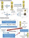
Clinical-Translational Model Incorporating Patient-Derived Xenografts. Two patient-derived xenograft (PDX) clinical-translational models are shown. The tumor avatar approach (Top panel) is one in which the patient's tumor is used to generate a PDX that will directly inform his or her own therapy. A schematic is shown for one potential tumor avatar design in which an individual patient and a PDX derived from this patient's own cancer are given standard of care (SOC) therapy, such as chemotherapy (chemo) and radiation (XRT). When the PDX develops resistance to the SOC, the resistant PDX is then expanded and tested against potential second line therapies (Tx1-4). The winning therapy (indicated by trophy) is then selected for second line therapy for the patient when they develop tumor recurrence following SOC. A tumor proband approach is shown schematically (bottom panel). In a proband model system, a cohort of PDX are utilized that represent the spectrum of human disease for that tumor type. These PDX are molecularly profiled and tested against various therapeutic regimens, such as irradiation (XRT), chemotherapeutics, and small molecule inhibitors. Ideally, molecular profiling is performed longitudinally (i.e., pre- and post- therapy) as molecular profiles likely change due to tumor adaptation/response. Well-characterized PDX will generate a library of profiling and phenotype information that can be used to inform clinical decisions as follows: a patient's tumor is molecularly profiled and matched to a particular or multiple PDX and known therapeutic sensitivity information is then passed back to the clinician for selection of proband-directed therapy.

References

    1. Johnson JI, Decker S, Zaharevitz D, Rubinstein LV, Venditti JM, Schepartz S, Kalyandrug S, Christian M, Arbuck S, Hollingshead M, Sausville EA. Relationships between drug activity in NCI preclinical in vitro and in vivo models and early clinical trials. Br J Cancer. 2001;84:1424–1431. - PMC - PubMed
    1. Williams SA, Anderson WC, Santaguida MT, Dylla SJ. Patient-derived xenografts, the cancer stem cell paradigm, and cancer pathobiology in the 21st century. Lab Invest. 2013;93:970–982. - PubMed
    1. Choi SY, Lin D, Gout PW, Collins CC, Xu Y, Wang Y. Lessons from patient-derived xenografts for better in vitro modeling of human cancer. Adv Drug Deliv Rev. 2014;79-80C:222–237. - PubMed
    1. Bredel M, Jacoby E. Chemogenomics: an emerging strategy for rapid target and drug discovery. Nat Rev Genet. 2004;5:262–275. - PubMed
    1. Gerlinger M, Rowan AJ, Horswell S, Larkin J, Endesfelder D, Gronroos E, Martinez P, Matthews N, Stewart A, Tarpey P, Varela I, Phillimore B, Begum S, McDonald NQ, Butler A, Jones D, Raine K, Latimer C, Santos CR, Nohadani M, Eklund AC, Spencer-Dene B, Clark G, Pickering L, Stamp G, Gore M, Szallasi Z, Downward J, Futreal PA, Swanton C. Intratumor heterogeneity and branched evolution revealed by multiregion sequencing. N Engl J Med. 2012;366:883–892. - PMC - PubMed

Publication types