. 2015 Oct 6;6(30):28539-55.
doi: 10.18632/oncotarget.5164.

A novel human truncated IL12rβ1-Fc fusion protein ameliorates experimental autoimmune encephalomyelitis via specific binding of p40 to inhibit Th1 and Th17 cell differentiation

Affiliations

A novel human truncated IL12rβ1-Fc fusion protein ameliorates experimental autoimmune encephalomyelitis via specific binding of p40 to inhibit Th1 and Th17 cell differentiation

Wei Guo et al. Oncotarget. .

Abstract

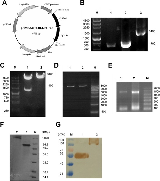
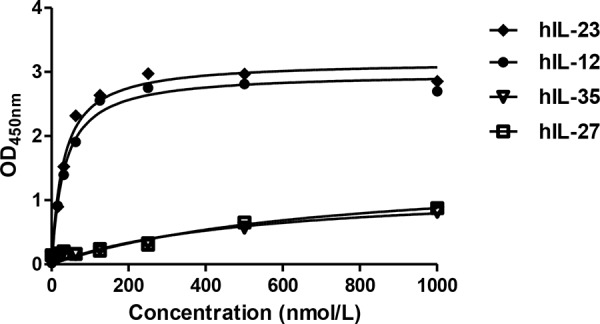
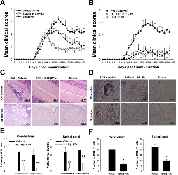
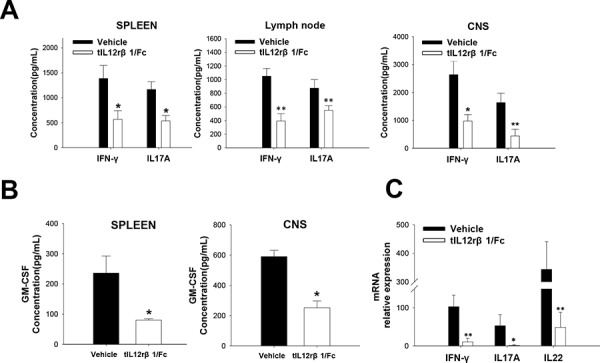
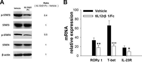
Interleukin (IL)-12 and IL-23 respectively driving polarization of T helper (Th) 1 and Th17 cells has been strongly implicated in the pathogenesis of both multiple sclerosis (MS) and experimental autoimmune encephalomyelitis (EAE). In this study, we first constructed, expressed and purified a novel human truncated IL12rβ1-Fc fusion protein (tIL12rβ1/Fc) binding multiple forms of the p40 subunit of human IL-12 and IL-23. tIL12rβ1/Fc was found to effectively ameliorate MOG35-55-induced EAE through reducing the production of Th1- and Th17-polarized pro-inflammatory cytokines and suppressing inflammation and demyelination in the focused parts. Moreover, tIL12rβ1/Fc suppressed Th1 (IFN-γ(+) alone) and IFN-γ(+) IL-17(+) as well as the population of classic Th17 (IL-17(+) alone) cells in vivo. Furthermore, tIL12rβ1/Fc ameliorated EAE at the peak of disease via the inhibition of STAT pathway, thereby causing a prominent reduction of RORγt (Th17) and T-bet (Th1) expression. Notably, tIL12rβ1/Fc could increase the relative number of CD4(+) Foxp3(+) regulatory T cells. These findings indicates that tIL12rβ1/Fc is a novel fusion protein for specific binding multiple forms of p40 subunit to exert potent anti-inflammatory effects and provides a valuable approach for the treatment of MS and other autoimmune diseases.

Keywords: Immune response; Immunity; Immunology and Microbiology Section; autoimmune disease; binding specificity; central nervous system; cytokine-blocking; molecular mechanism.


Conflict of interest statement

CONFLICTS OF INTEREST

The authors declare that they have no competing interests.

Figures

Figure 1
Figure 1. Construction, expression, and purification of tIL12rβ1/Fc
A. Plasmid map of the eukaryotic expression plasmid containing human truncated IL-12rβ1 receptor (pcDNA3.1(+)-tIL12rβ1/Fc). The gene sequence encoding tIL12rβ1/Fc was inserted into pcDNA3.1(+) vector at the corresponding restriction sites Hind III and Xho I. B. PCR products of tIL12rβ1/Fc recombinant gene. Lane 1, tIL12rβ1/Fc gene sequence containing the signal peptide. Lane 2, IgG1 Fc fragment. Lane 3, recombinant tIL12rβ1/Fc fused gene product using overlap PCR. M, DNA molecular weight markers, bp. C. Correctly constructed plasmid was verified by digestion at the Hind III and Xho I sites. Lane 1, pcDNA3.1(+)-tIL12rβ1/Fc after digestion. Lane 2, pcDNA3.1(+) -tIL12rβ1/Fc before digestion. M, DNA molecular weight markers, bp. D. Plasmids were linearized for cell transfection using Pvu I. Lane 1 and 2, linearized pcDNA3.1(+)-tIL12rβ1/Fc. M, DNA molecular weight markers, bp. E. Total RNA of the two strains were identified by RT-PCR with primers F1 and F4. F. SDS-PAGE analysis of the purified tIL12rβ1/Fc fusion protein using Protein A column. Lane 1, purified tIL12rβ1/Fc fusion protein at reduced state. Lane 2, purified tIL12rβ1/Fc fusion protein at non-reduced state. M, protein molecular weight markers, KDa. G. Western blot analysis of tIL12rβ1/Fc using mAbs against human IL12rβ1. Lane 1, purified tIL12rβ1/Fc fusion protein at reduced state. Lane 2, purified tIL12rβ1/Fc fusion protein at non-reduced state. M, protein molecular weight markers, KDa.
Figure 2
Figure 2. Binding affinity of tIL12rβ1/Fc protein to hIL-12/IL-23 and hIL -27/IL-35
Binding affinity of tIL12rβ1/Fc protein to human IL-12 and human IL-23 were examined by direct binding ELISA. Meanwhile, the binding assays of tIL12rβ1/Fc protein to human IL-27 and IL-35 were also conducted as controls to confirm the binding specificity, showing no cross-reactions with our molecule.
Figure 3
Figure 3. Amelioration of EAE by tIL12rβ1/Fc treatment
The tIL12rβ1/Fc or vehicle (saline) was administered via tail vein injection at 2.5 mg/kg and CsA was administered i.g. as positive control at 1.5 mg/kg. The regimen started 9 days after immunization as therapeutic regimen (A) or 3 days before immunization as preventive regimen (B). C. H&E staining analysis of brains and spinal cords obtained from normal mice or mice treated with vehicle or tIL12rβ1/Fc at day 30 post-immunization (therapeutic protocol). D. Immunohistochemistry for CD4+ T cell infiltration of brains and spinal cords obtained from normal mice or mice treated with vehicle or tIL12rβ1/Fc at day 30 post-immunization (therapeutic protocol). E. Pathology scores of inflammation and demyelination in brains and spinal cords. F. The number of CD4+ T cells in spinal cords and brains. Data are expressed as mean ± SD (n = 9). *p < 0.05; **p < 0.01.
Figure 4
Figure 4. Downregulation of Th1 and Th17 cells by tIL12rβ1/Fc treatment
Splenocytes, lymph nodes cells and CNS MNCs of tIL12rβ1/Fc- or vehicle-treated EAE mice at day 18 post-immunization (therapeutic protocol) were isolated. A. Supernatants derived from splenocytes, lymph nodes cells and CNS MNCs reactivated with MOG35–55 (20 μg/mL) for 24 h were analyzed for the level of p40 (mean ± SD; n = 5). B. The percentage of Th1, Th1/Th17 and Th17 cells in the CD4+ lymphocyte gate were analyzed by intracellular staining of IFN-γ and IL-17, following stimulation with MOG35–55 (20 μg/mL) for 24 h. C. The Th17:Th1 ratios in CNS, LNs and Spleens were observed for each group. Correlation of Th17:Th1 ratios with vehicle treatment and that with tIL12rβ1/Fc treatment was significant (p < 0.05) (n = 4). D. Percentages of cells positive expression with these antigens in spleen, LN or CNS are expressed as mean ± SD (n = 4). E. The frequency of regulatory T cells in the CD4+ lymphocyte gate were analyzed by intracellular staining of Foxp3 following stimulation with MOG35–55 (20 μg/mL) for 24 h. Percentage of Foxp3+ cells in spleen is expressed as mean ± SD (n = 4). *p < 0.05; **p < 0.01; ***p < 0.001.
Figure 5
Figure 5. Suppression of related cytokines by tIL12rβ1/Fc treatment
Splenocytes, lymph nodes cells and CNS MNCs of tIL12rβ1/Fc- or vehicle-treated EAE mice at day 18 post-immunization (therapeutic protocol) were isolated. A. Supernatants derived from splenocytes, lymph nodes cells and CNS MNCs reactivated with MOG35–55 (20 μg/mL) for 24 h were analyzed for the level of IFN-γ and IL-17A (mean ± SD; n = 5). B. Supernatants derived from splenocytes and CNS MNCs reactivated with MOG35–55 (20 μg/mL) for 24 h were also measured for the expression of GM-CSF. C. mRNA level of IFN-γ, IL-17A and IL-22 from the same CD4+ splenocyte preparations were analyzed by quantitative real-time PCR. Data are representative of three independent experiments. *p < 0.05; **p < 0.01.
Figure 6
Figure 6. Role of tIL12rβ1/Fc in mouse Th1 and Th17 cell differentiation
Mouse naive CD4+ cells were differentiated into Th1 or Th17 cells in the presence of different concentrations of tIL12rβ1/Fc in vitro. A. The percentage of Th1 and Th17 cells in the CD4 subset were analyzed by intracellular staining of IFN-γ and IL-17A, respectively. B. Supernatants derived from cultured spleen cells, as above, were analyzed for the indicated cytokines. C. mRNA abundance of transcription factor RORγt and T-bet were also measured by quantitative real-time RT-PCR. Data are representative of three independent experiments. *p < 0.05; **p < 0.01; ***p < 0.001.
Figure 7
Figure 7. Role of tIL12rβ1/Fc in human Th1 and Th17 cell differentiation
Human PBMCs purified by density gradient centrifugation using Histopaque were differentiated into Th1 or Th17 cells in the presence of indicated tIL12rβ1/Fc. A. Human Th1 and Th17 development in the CD4 gate in PBMCs were examined by Flow cytometry analysis. B. The level of secreted IFN-γ and IL-17A in cell culture supernatants were analyzed using ELISA method. C. mRNA expression of T-bet and RORγt from the same cell preparations were analyzed by quantitative real-time PCR. Data are representative of three independent experiments. **p < 0.01; ***p < 0.001.
Figure 8
Figure 8. The inhibitory effect of tIL12rβ1/Fc on Th1 and Th17 in relation to STAT signaling pathway
A. Purified CD4+ splenocytes from tIL12rβ1/Fc-treated mice or control EAE mice on day 18 post-immunization (therapeutic protocol) were analyzed by Western blot assay for the expression or phosphorylation of indicated proteins in the STAT pathway. B. The same CD4+ T cell preparations were measured for the mRNA abundance of RORγt, T-bet and IL-23R by quantitative real-time RT-PCR. Data are representative of three independent experiments. *p < 0.05; **p < 0.01; ***p < 0.001.
Figure 9
Figure 9. Inhibition of NF-κB signaling pathway by tIL12rβ1/Fc treatment
A. Splenocytes from tIL12rβ1/Fc or vehicle-treated EAE mice on day 18 post-immunization (therapeutic protocol) were analyzed for phosphorylated IκBα, phosphorylated p65, IκBα and p65 by Western blot assay. B. Expression levels of NF-κB-regulated gene products including IL-6, TNF-α and iNOS were measured by quantitative real-time RT-PCR. Data are representative of three independent experiments. **p < 0.01; ***p < 0.001.

Similar articles

Cited by

References

    1. Lu Y, Chen B, Song J-H, Zhen T, Wang B-Y, Li X, Liu P, Yang X, Zhang Q-L, Xi X-D. Eriocalyxin B ameliorates experimental autoimmune encephalomyelitis by suppressing Th1 and Th17 cells. Proceedings of the National Academy of Sciences. 2013;110:2258–2263. - PMC - PubMed
    1. Constantinescu CS, Farooqi N, O'Brien K, Gran B. Experimental autoimmune encephalomyelitis (EAE) as a model for multiple sclerosis (MS) British Journal of Pharmacology. 2011;164 - PMC - PubMed
    1. Costa GL, Sandora MR, Nakajima A, Nguyen EV, Taylor-Edwards C, Slavin AJ, Contag CH, Fathman CG, Benson JM. Adoptive immunotherapy of experimental autoimmune encephalomyelitis via T cell delivery of the IL-12 p40 subunit. The Journal of Immunology. 2001;167:2379–2387. - PubMed
    1. Moldovan IR, Rudick RA, Cotleur AC, Born SE, Lee J-C, Karafa MT, Pelfrey CM. Interferon gamma responses to myelin peptides in multiple sclerosis correlate with a new clinical measure of disease progression. Journal of neuroimmunology. 2003;141:132–140. - PubMed
    1. Steinman L. A brief history of T(H)17, the first major revision in the T(H)1/T(H)2 hypothesis of T cell-mediated tissue damage. Nature medicine. 2007;13:139–145. - PubMed

Publication types

MeSH terms