

Endothelial-derived hyperpolarization contributes to acetylcholine-mediated vasodilation in human skin in a dose-dependent manner
- PMID: 26384409
- PMCID: PMC4628993
- DOI: 10.1152/japplphysiol.00201.2015
Endothelial-derived hyperpolarization contributes to acetylcholine-mediated vasodilation in human skin in a dose-dependent manner
Abstract
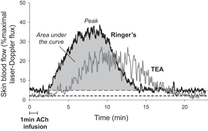
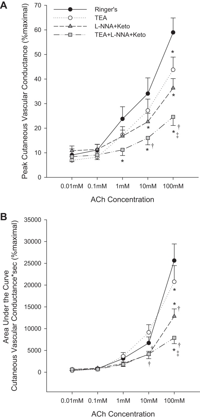
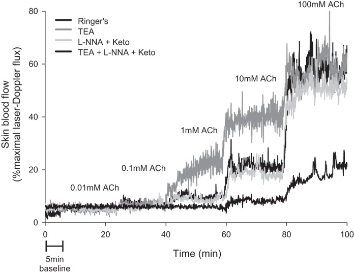
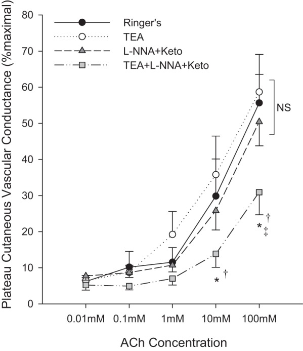
Cutaneous acetylcholine (ACh)-mediated dilation is commonly used to assess microvascular function, but the mechanisms of dilation are poorly understood. Depending on dose and method of administration, nitric oxide (NO) and prostanoids are involved to varying extents and the roles of endothelial-derived hyperpolarizing factors (EDHFs) are unclear. In the present study, five incremental doses of ACh (0.01-100 mM) were delivered either as a 1-min bolus (protocol 1, n = 12) or as a ≥20-min continuous infusion (protocol 2, n = 10) via microdialysis fibers infused with 1) lactated Ringer, 2) tetraethylammonium (TEA) [a calcium-activated potassium channel (KCa) and EDHF inhibitor], 3) L-NNA+ketorolac [NO synthase (NOS) and cyclooxygenase (COX) inhibitors], and 4) TEA+L-NNA+Ketorolac. The hyperemic response was characterized as peak and area under the curve (AUC) cutaneous vascular conductance (CVC) for bolus infusions or plateau CVC for continuous infusions, and reported as %maximal CVC. In protocol 1, TEA, alone and combined with NOS+COX inhibition, attenuated peak CVC (100 mM Ringer 59 ± 6% vs. TEA 43 ± 5%, P < 0.05; L-NNA+ketorolac 35 ± 4% vs. TEA+L-NNA+ketorolac 25 ± 4%, P < 0.05) and AUC (Ringer 25,414 ± 3,528 vs. TEA 21,403 ± 3,416%·s, P < 0.05; L-NNA+ketorolac 25,628 ± 3,828%(.)s vs. TEA+L-NNA+ketorolac 20,772 ± 3,711%·s, P < 0.05), although these effects were only significant at the highest dose of ACh. At lower doses, TEA lengthened the total time of the hyperemic response (10 mM Ringer 609 ± 78 s vs. TEA 860 ± 67 s, P < 0.05). In protocol 2, TEA alone did not affect plateau CVC, but attenuated plateau in combination with NOS+COX inhibition (100 mM 50.4 ± 6.6% vs. 30.9 ± 6.3%, P < 0.05). Therefore, EDHFs contribute to cutaneous ACh-mediated dilation, but their relative contribution is altered by the dose and infusion procedure.
Keywords: calcium-activated potassium channels; endothelium; laser-Doppler flowmetry; nitric oxide; skin.
Copyright © 2015 the American Physiological Society.
Figures
References
-
- Agarwal SC, Allen J, Murray A, Purcell IF. Laser Doppler assessment of dermal circulatory changes in people with coronary artery disease. Microvasc Res 84: 55–59. - PubMed
-
- Ay I, Tuncer M. Both endothelium and afferent nerve endings play a role in acetylcholine-induced renal vasodilation. Life Sci 79: 877–882, 2006. - PubMed
-
- Bauersachs J, Popp R, Hecker M, Sauer E, Fleming I, Busse R. Nitric oxide attenuates the release of endothelium-derived hyperpolarizing factor. Circulation 94: 3341–3347, 1996. - PubMed
-
- Berghoff M, Kathpal M, Kilo S, Hilz MJ, Freeman R. Vascular and neural mechanisms of ACh-mediated vasodilation in the forearm cutaneous microcirculation. J Appl Physiol 92: 780–788, 2002. - PubMed
-
- Brayden JE. Membrane hyperpolarization is a mechanism of endothelium-dependent cerebral vasodilation. Am J Physiol Heart Circ Physiol 259: H668–H673, 1990. - PubMed
Publication types
MeSH terms
Substances
Grants and funding
LinkOut - more resources
Full Text Sources
Other Literature Sources