. 2016 Jan 8;44(1):152-63.
doi: 10.1093/nar/gkv900. Epub 2015 Sep 17.

Guanine quadruplex structures localize to heterochromatin

Affiliations

Guanine quadruplex structures localize to heterochromatin

Roland F Hoffmann et al. Nucleic Acids Res. .

Erratum in

  • Guanine quadruplex structures localize to heterochromatin.
    Hoffmann RF, Moshkin YM, Mouton S, Grzeschik NA, Kalicharan RD, Kuipers J, Wolters AHG, Nishida K, Romashchenko AV, Postberg J, Lipps H, Berezikov E, Sibon OCM, Giepmans BNG, Lansdorp PM. Hoffmann RF, et al. Nucleic Acids Res. 2017 Jun 2;45(10):6253. doi: 10.1093/nar/gkx301. Nucleic Acids Res. 2017. PMID: 28449026 Free PMC article. No abstract available.

Abstract

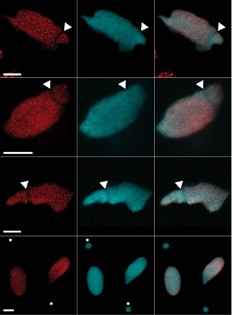
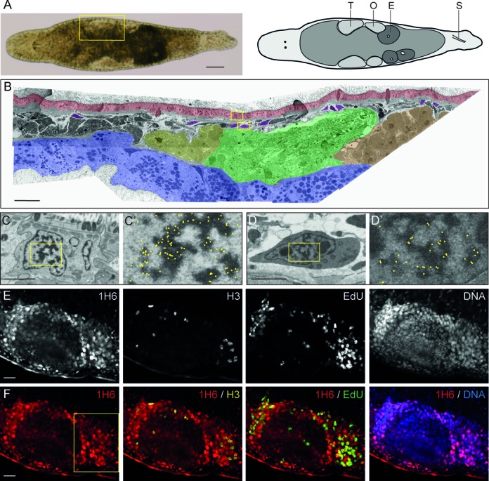
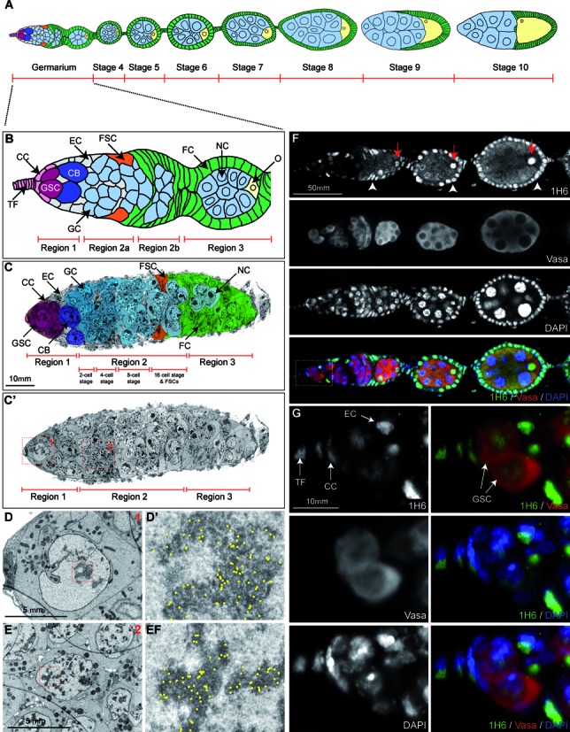
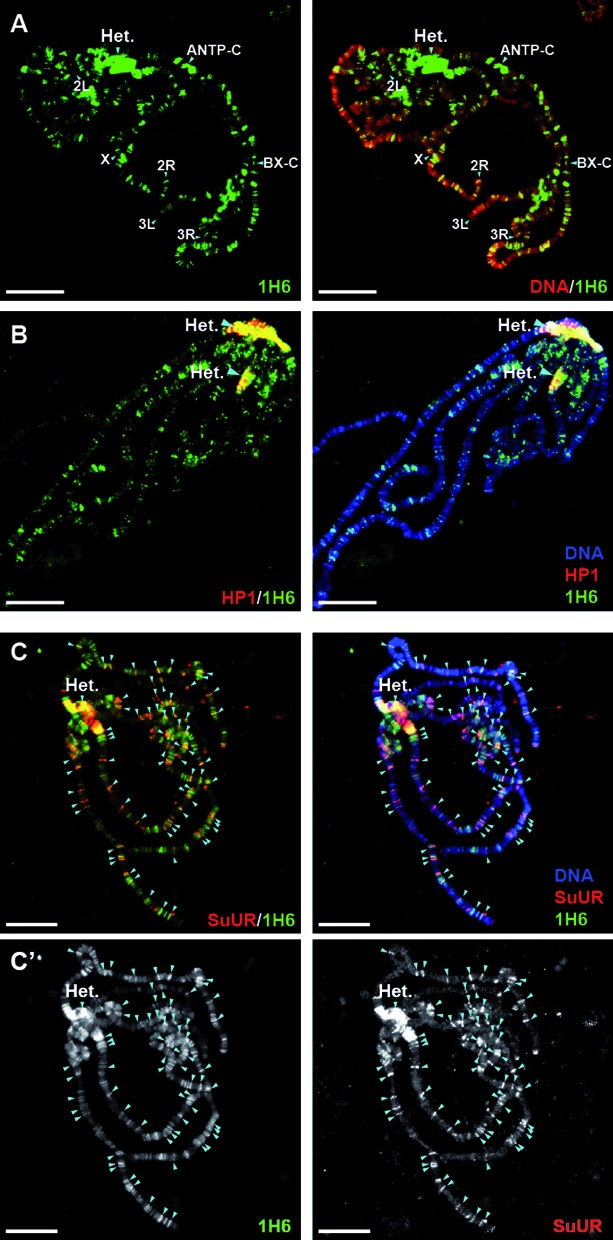
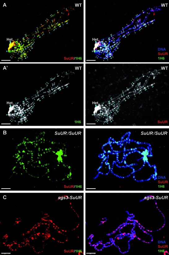
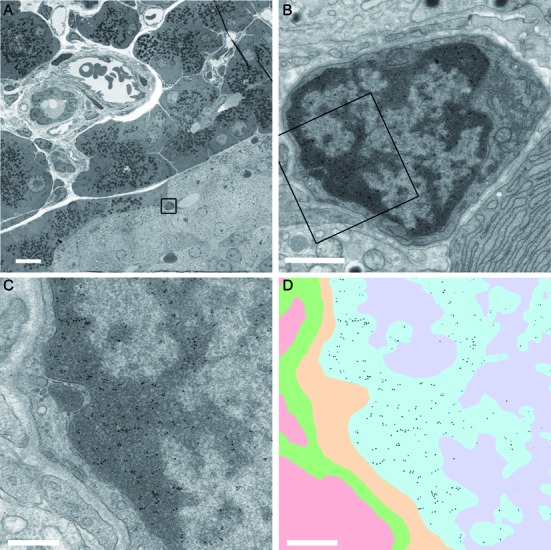
Increasing amounts of data support a role for guanine quadruplex (G4) DNA and RNA structures in various cellular processes. We stained different organisms with monoclonal antibody 1H6 specific for G4 DNA. Strikingly, immuno-electron microscopy showed exquisite specificity for heterochromatin. Polytene chromosomes from Drosophila salivary glands showed bands that co-localized with heterochromatin proteins HP1 and the SNF2 domain-containing protein SUUR. Staining was retained in SUUR knock-out mutants but lost upon overexpression of SUUR. Somatic cells in Macrostomum lignano were strongly labeled, but pluripotent stem cells labeled weakly. Similarly, germline stem cells in Drosophila ovaries were weakly labeled compared to most other cells. The unexpected presence of G4 structures in heterochromatin and the difference in G4 staining between somatic cells and stem cells with germline DNA in ciliates, flatworms, flies and mammals point to a conserved role for G4 structures in nuclear organization and cellular differentiation.


Figures

Figure 1.
Figure 1.
Staining of G4 DNA in the macronucleus of Stylonychia lemnea. Indirect immunofluorescence of Stylonychia using monoclonal antibody 1H6. Shown are different adult animals with a characteristic staining pattern that excludes the replication band (arrows) as well as the micronucleus (bottom panels, asterisks). The left panels show 1H6 staining, the second row corresponding DAPI staining and the third column an overlay of 1H6 and DAPI fluorescence. Scale bars: 10 μm.
Figure 2.
Figure 2.
Weak 1H6 staining of proliferating cells in adult Macrostomum. (A) Bright-field image and schematic overview of Macrostomum lignano. T: Testis, O: Ovary, E: Egg and S: Stylet. (B) False-colored overview of 1H6 immuno-EM nanotomy data of Figure 2 available at http://www.nanotomy.org/. Red: Epidermis; Blue: Gut; Yellow: Testis Tip; Green: Testis; Orange: Ovary; Purple: somatic stem cells (neoblasts). (C–D’) EM images of an epidermal cell (C, C’) and a neoblast (D, D’) following labeling with 1H6 antibody. Boxed areas in B are shown in C and D. Boxed area in C and D are shown in C’ and D’. For clarity, the immunogold particles are marked manually with yellow dots in C’ and D’ (raw data available online). (E) Gray scale images of a section through the germline. stained with 1H6, EdU (S-phase cells), phospho-H3 (mitotic cells) and DNA. (F) Colored overlays of sections shown in E. Higher magnifications of the boxed area in F are shown in Supplementary Figure S3. Scale bars: A: 100 μm, B: 30 μm; C, D: 2 μm; E–F: 20 μm.
Figure 3.
Figure 3.
Germline stem cells in Drosophila contain less heterochromatin and stain weaker with 1H6 compared to most other cells. (A) Schematic diagram of a Drosophila ovariole up to stage 10 of egg chamber development. Green: follicle cells; light blue: nurse cells; yellow: developing oocyte. (B) Schematic of the germarium. TF: Terminal Filament; CC = Cap Cells; GSC: Germline Stem Cell; CB: Cystoblasts; EC: Escort Cells; GC: Germline Cysts; FSC: Follicle Stem Cells; FC: Follicle Cells; NC: Nurse Cells; O: Oocyte. (C–E) Overview (C,C’) and close-up (D,E) images of a germarium, processed for EM and immunogold-labeled with 1H6 antibody. Nanotomy of Figure 2 shown in C’ is available online at http://www.nanotomy.org). (C,C’) Overview of the germarium up to stage 3. Individual cell types, identified to the best of our knowledge, were false-colored with Photoshop to match the schematic shown in B. Close-up snapshots shown in D and E correspond to the red-colored boxes in C’ and depict a Germline Stem Cell (D) and Germline Cyst Cell (E) and close-ups of their heterochromatic nuclear regions respectively (D’ and E’). Gold-particles, marking the presence of the 1H6-antibody, were manually marked with yellow dots (raw data available online) in D’ and E’. Note the co-localization with heterochromatin. (F and G) Part of a Drosophila ovariole (germarium and stage 4–5, compare with schematic in A, stained with anti-1H6 (green), anti-Vasa, to indicate the germline cells (red) and the DNA dye DAPI (blue). Higher magnification images in G correspond to the white box shown in F. In the Drosophila germarium 1H6 antibody shows strong preference for the Terminal Filament, Cap Cells, Escort Cells (indicated in G), Follicle Cells (white arrow heads in F) and the developing oocyte (red arrows in F). This can also be seen in later stages of egg development (Supplementary Figure S2). In contrast, the Vasa-positive Germline Stem Cells, Germline Cysts and future Nurse Cells show much weaker 1H6 staining. Scale bars: C: 10 μm; D–E: 5 μm; F: 50 μm and G: 10 μm.
Figure 4.
Figure 4.
1H6 staining of polytene chromosomes. (A) Chromosomes from salivary glands of third instar Drosophila larvae, stained for 1H6 (green) and the DNA dye DAPI (red). Drosophila salivary glands undergo through multiple rounds of endoreplication, resulting in polytene chromosomes that can easily be visualized with antibody staining and/or dyes. 1H6 staining reveals multiple specific loci on polytene chromosomes including pericentric heterochromatin (Het.) and developmental loci harboring Antennapedia (ANTP-C) and Bithorax Complex (BX-C). The regions of ANTP-C and BX-C, as well as pericentric heterochromatin are late replicated and underreplicated in polytene chromosomes (57). The tips of chromosome arms X, 2L, 2R, 3L and 3R are labeled. Scale bar: 25 μm. (B) Pericentric heterochromatin is enriched in DNA quadruplexes. Polytene chromosomes were stained with antibodies raised against Heterochromatin Protein 1 (HP1, red) and G4 DNA (1H6, green). Pericentric heterochromatin (Het.) is strongly marked with both antibodies appearing yellow when merged (right panel). Scale bar: 25 μm. (C–C’) G4 DNA co-localizes with the regions of late replication marked by SUUR. Polytene chromsomes were stained with SUUR (red) and 1H6 (green). The bands, which are labeled by both 1H6 and anti-SUUR antibodies are indicated by arrowheads. Note that, although most of the SUUR binding sites are also stained by 1H6, there are more additional loci stained only by 1H6. (C’) Split, black and white images for 1H6 (left panel) and SUUR (right panel). Scale bar: 25 μm.
Figure 5.
Figure 5.
Loss of G4 structures upon overexpression of SUUR. (A–A’) In polytene chromosomes of wild type third instar Drosophila larvae, nearly all SUUR binding loci co-localize with G4 DNA (arrowheads) labeled by anti-SUUR (red) and 1H6 (green) antibodies. (A’) Split, black and white images for 1H6 (left panel) and SUUR (right panel). (B) In polytene chromsomes of larvae homozygous for SUUR mutant allele G4 DNA loci are retained. Polytene chromsomes were stained with SUUR (red) and 1H6 (green). Note that SUUR staining is absent. (C) In polytene chromosomes of larvae overexpressing SUUR under the control of salivary gland specific GAL4 driver (sgs3), 1H6 staining is lost. Polytene chromosomes were stained with SUUR (red) and 1H6 (green). Note the increased intensity of SUUR staining. Scale bar: 25 μm.
Figure 6.
Figure 6.
1H6 binds to heterochromatin in different cells of rat pancreas. (A) Overview of pancreatic tissue with alpha cells, beta cells, endothelial cells, epithelial cells and exocrine cells. The different cell types can be recognized as described (www.nanotomy.nl) (27). A high-resolution ‘nanotomy’ digital file of Figure 6, were gold-particles can be detected in a ‘Google-earth’ like analysis, is available online at http://www.nanotomy.org/). The boxed area in (A) is shown in (B). The boxed area in (B) is shown in (C) and (D). Annotation of the ultrastructure (C) is illustrated in (D) as follows: heterochromatin blue; euchromatin purple; cytoplasm yellow; extracellular space in green and an adjacent cell (cytoplasm) in pink. The data of panel (C) were processed to selectively visualize the gold particles (black). Bars: A: 10 μm; B: 1 μm; C,D: 0.5 μm.

Similar articles

Cited by

References

    1. Jost K.L., Bertulat B., Cardoso M.C. Heterochromatin and gene positioning: inside, outside, any side. Chromosoma. 2012;121:555–563. - PMC - PubMed
    1. Passarge E. Emil Heitz and the concept of heterochromatin: longitudinal chromosome differentiation was recognized fifty years ago. Am. J. Hum. Genet. 1979;31:106–115. - PMC - PubMed
    1. Woodcock C.L., Ghosh R.P. Chromatin higher-order structure and dynamics. Cold Spring Harb. Perspect. Biol. 2010;2:a000596. - PMC - PubMed
    1. Bickmore W.A., van Steensel B. Genome architecture: domain organization of interphase chromosomes. Cell. 2013;152:1270–1284. - PubMed
    1. Hubner M.R., Eckersley-Maslin M.A., Spector D.L. Chromatin organization and transcriptional regulation. Curr. Opin. Genet. Dev. 2013;23:89–95. - PMC - PubMed

Publication types