. 2015 Oct 15;43(18):8615-26.
doi: 10.1093/nar/gkv909. Epub 2015 Sep 17.

Structure of a human translation termination complex

Affiliations

Structure of a human translation termination complex

Sarah Matheisl et al. Nucleic Acids Res. .

Abstract

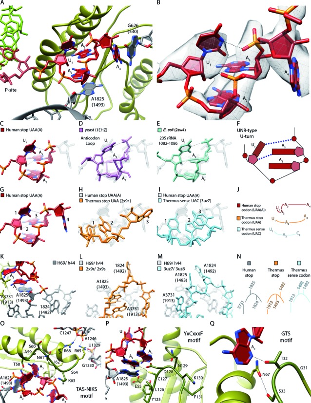
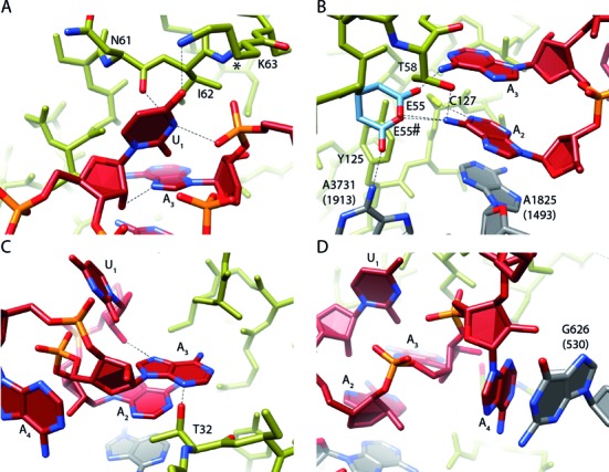
In contrast to bacteria that have two release factors, RF1 and RF2, eukaryotes only possess one unrelated release factor eRF1, which recognizes all three stop codons of the mRNA and hydrolyses the peptidyl-tRNA bond. While the molecular basis for bacterial termination has been elucidated, high-resolution structures of eukaryotic termination complexes have been lacking. Here we present a 3.8 Å structure of a human translation termination complex with eRF1 decoding a UAA(A) stop codon. The complex was formed using the human cytomegalovirus (hCMV) stalling peptide, which perturbs the peptidyltransferase center (PTC) to silence the hydrolysis activity of eRF1. Moreover, unlike sense codons or bacterial stop codons, the UAA stop codon adopts a U-turn-like conformation within a pocket formed by eRF1 and the ribosome. Inducing the U-turn conformation for stop codon recognition rationalizes how decoding by eRF1 includes monitoring geometry in order to discriminate against sense codons.


Figures

Figure 1.
Figure 1.
Translation in human cell-free extract. (A) Schematic representation of the individual steps from human HeLa cell culture to a cell-free human in vitro translation extract. Cells were grown in a large spinner flask. After harvesting and washing of the cells, they were subjected to nitrogen pressure and disrupted by sudden pressure release. A quick centrifugation step resulted in a crude extract which can be supplemented for optimal performance of mRNA in vitro translation. Example of in vitro translational stalling optimization for (B) time (C) magnesium acetate concentration and (D) potassium acetate concentration using Western blotting with anti-HA antibody for detection of tRNA-bound hCMV peptide or free hCMV peptide. Optimal conditions vary for each extract.
Figure 2.
Figure 2.
Isolation and cryo-EM structure of a stalled human 80S ribosome bound to eRF1. (A) Schematic representation of the hCMV stalling mRNA construct used in the human translation system for 80S-CMV-RNC generation. The final mRNA construct encoded a CrPV IGR IRES sequence for translation initiation, an N-terminal HA- and (His)6-tag, the well characterized DP75 dipeptidyl-aminopeptidase B (DPAPB) (30), the hCMV uORF2 stalling sequence and a polyA-tail. The termination site relevant for the Stop23Ala mutated construct is underlined. (B, C) Western blots of purified human RNCs programmed with the hCMV mRNA shown in (A) or programmed with truncated mRNA (harbouring no stop codon in the A-site). Signal detection was performed with anti-HA antibody for detection of (B) peptidyl-tRNA or (C) anti-eRF1 antibody. (D) Cryo-EM structure of the eRF1-bound human 80S-CMV-RNC at 3.8 Å resolution. The colour code for mRNA, tRNA and the eRF1 domains is given. (E) Section of (D) focusing on the hCMV peptidyl-tRNA. (F) Top view section of the human eRF1 bound 80S-CMV-RNC cryo-EM structure. (G) Molecular models of (F). For docking, the model of the human ribosome POST structure (pdb code: 5AJ0) and the crystal/NMR structures of human eRF1 (pdb codes: 3E1Y-A, 2KTV) were used. (H, I) Molecular models displaying (H) all 3 eRF1 domains and (I) ribosomal contacts of human eRF1.
Figure 3.
Figure 3.
Mechanism of termination silencing by nascent hCMV peptide. (A) Schematic representation of the gp48/UL4 mRNA illustrating expression regulation of gp48/UL4 by leaky scanning of uORF2. (B) Ribosome stalling on the hCMV uORF2 leads to gp48/UL4 repression. (C) Identification of arrest-defective amino acid substitutions within the hCMV stalling peptide indicated by decreased amount of hCMV peptidyl-tRNA and accumulation of free peptide detected by Western blotting. (D, upper left) Overview of eRF1, peptidyl-tRNA and ribosomal proteins uL22 and uL4 labelled and coloured distinctively. (D, lower left) EM density section revealing the helical structure which is formed by the nascent hCMV peptide within the upper part of the ribosomal tunnel. (D, right) Same section, showing H133 of uL22 and nucleotides of the ribosomal tunnel wall which interact with the helical part of the nascent chain (arrows). (E) Position of the eRF1 GGQ-motif and the peptidyl-tRNA within the PTCs. (F) Comparison of the position of the bacterial RF2 and the human eRF1 GGQ-motifs and the peptidyl-tRNAs within the PTC. (G) eRF1-stabilizing position of Hs A4510 (Ec A2602). (H-K) Comparison of the position of Hs U4493 (Ec U2585) in (H) the hCMV stalled ribosome complex, (I) the human ribosome in the POST state, (J) the prokaryotic pre-termination complex and (K) the ErmCL stalled ribosomal complex.
Figure 4.
Figure 4.
U-turn-like geometry of the UAA stop-codon bound by eRF1 and the ribosome. (A) Human UAA(A) stop-codon (red) positioned in a cavity between eRF1 (green) and the ribosomal bases Hs G626 (Ec G530) and Hs A1825 (Ec A1493) (gray). (B) EM density revealing the geometry of the mRNA sequence UAA(A) during stop codon recognition. (C–F) Comparison of the human stop codon mRNA geometry with the known UNR-type U-turn structures (D) yeast anticodon loop (purple) and (E) Ec 23S rRNA (1082–1086) (turquois). (F) Schematic representation of (CE). (G–J) Comparison of the A-site mRNA geometry during (G) human UAA stop codon recognition (H), Thermus thermophilus (Tt) UAA stop codon recognition (light orange) and (I) Tt UAC sense decoding (light blue). (J) Schematic representation of (GI). (K–N) Comparison of the h44 (Hs A1824 (Ec A1492), Hs A1825 (Ec A1493))/H69 (Hs A3731 (Ec A1913)) states during (K) human UAA stop codon recognition, (L) Tt UAA stop codon recognition and (M) Tt UAC decoding. (N) Schematic representation of (KM). (O) Interactions of the eRF1 TAS-NIKS motif (residues 58–64) with the mRNA/rRNA. The hydroxylation site of K63 is located at C4 (*). (P) Interactions of the eRF1 YxCxxxF (residues 125–131) motif with the mRNA/rRNA. (Q) Interactions of the eRF1 GTS (residues 31–33) motif with the mRNA.
Figure 5.
Figure 5.
Interaction of eRF1 with the UAA(A) stop codon. (A) The Uracil 1 (U1) nucleotide of the stop codon interacts with N7 of adenosine 3 (A3) and the A3 backbone phosphate to adopt U-turn geometry of the stop codon. Further, U1 interacts with N61 and K63 of the eRF1 TAS-NIKS motif. The hydroxylation site of K63 is located at C4 (*). (B) Adenosine 2 (A2) interacts with Hs A1825 (h44) and C127 (YxCxxxF motif). Possible rotamer conformations of E55 (E55 and E55#) for the interaction with A2 are depicted in light blue. Y125 (YxCxxxF motif) stabilizes the shifted conformation of Hs A3731 (H69). (C) A3 mainly interacts with T32 (GTS motif) and the 2′OH of U1. (D) Adenosine 4 (A4) stacks on the rRNA base Hs G626 (Ec G530). Interactions are indicated by dotted lines.

References

    1. Ogle J.M., Carter A.P., Ramakrishnan V. Insights into the decoding mechanism from recent ribosome structures. Trends Biochem. Sci. 2003;28:259–266. - PubMed
    1. Demeshkina N., Jenner L., Westhof E., Yusupov M., Yusupova G. A new understanding of the decoding principle on the ribosome. Nature. 2012;484:256–259. - PubMed
    1. Zhou J., Korostelev A., Lancaster L., Noller H.F. Crystal structures of 70S ribosomes bound to release factors RF1, RF2 and RF3. Curr. Opin. Struct. Biol. 2012;22:733–742. - PMC - PubMed
    1. Korostelev A.A. Structural aspects of translation termination on the ribosome. RNA. 2011;17:1409–1421. - PMC - PubMed
    1. Rodnina M.V. The ribosome as a versatile catalyst: reactions at the peptidyl transferase center. Curr. Opin. Struct. Biol. 2013;23:595–602. - PubMed

Publication types

LinkOut - more resources