. 2015 Dec;24(12):1979-96.
doi: 10.1002/pro.2807. Epub 2015 Oct 4.

NMR analysis of the interaction of picornaviral proteinases Lb and 2A with their substrate eukaryotic initiation factor 4GII

Affiliations

NMR analysis of the interaction of picornaviral proteinases Lb and 2A with their substrate eukaryotic initiation factor 4GII

Martina Aumayr et al. Protein Sci. 2015 Dec.

Abstract

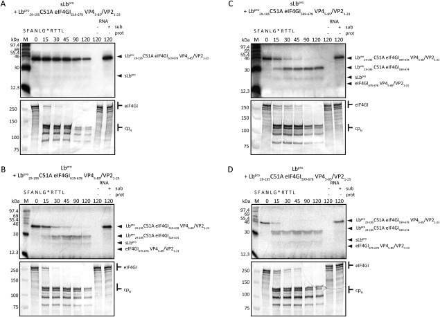
Messenger RNA is recruited to the eukaryotic ribosome by a complex including the eukaryotic initiation factor (eIF) 4E (the cap-binding protein), the scaffold protein eIF4G and the RNA helicase eIF4A. To shut-off host-cell protein synthesis, eIF4G is cleaved during picornaviral infection by a virally encoded proteinase; the structural basis of this reaction and its stimulation by eIF4E is unclear. We have structurally and biochemically investigated the interaction of purified foot-and-mouth disease virus (FMDV) leader proteinase (Lb(pro)), human rhinovirus 2 (HRV2) 2A proteinase (2A(pro)) and coxsackievirus B4 (CVB4) 2A(pro) with purified eIF4GII, eIF4E and the eIF4GII/eIF4E complex. Using nuclear magnetic resonance (NMR), we completed (13)C/(15) N sequential backbone assignment of human eIF4GII residues 551-745 and examined their binding to murine eIF4E. eIF4GII551-745 is intrinsically unstructured and remains so when bound to eIF4E. NMR and biophysical techniques for determining stoichiometry and binding constants revealed that the papain-like Lb(pro) only forms a stable complex with eIF4GII(551-745) in the presence of eIF4E, with KD values in the low nanomolar range; Lb(pro) contacts both eIF4GII and eIF4E. Furthermore, the unrelated chymotrypsin-like 2A(pro) from HRV2 and CVB4 also build a stable complex with eIF4GII/eIF4E, but with K(D) values in the low micromolar range. The HRV2 enzyme also forms a stable complex with eIF4E; however, none of the proteinases tested complex stably with eIF4GII alone. Thus, these three picornaviral proteinases have independently evolved to establish distinct triangular heterotrimeric protein complexes that may actively target ribosomes involved in mRNA recruitment to ensure efficient host cell shut-off.

Keywords: control of protein synthesis; protein-protein interactions; substrate binding; viral proteases; virus-host interactions.


Figures

Figure 1
Figure 1
Domain structure of human eIF4GII and cleavage of eIF4GII551–745 by picornaviral proteases. (A) Schematic representation of human eIF4G with known structural domains indicated (the “ring” domain has only been shown in yeast) and the amino acid sequence of the fragment of eIF4GII551–745 used. Protein binding sites are indicated as follows: eIF4E binding site, broken line; Lbpro binding site, dotted line; 2Apro binding region, straight line. Positions of the cleavage site of the Lbpro and 2Apro are marked by black and white triangles, respectively. The backbone NMR signals of 178 residues underlaid in black were assigned using triple resonance experiments, (B) In vitro cleavage of 5 µM eIF4GII551–745 by 7 nM sLbpro in the absence or the presence of 5 µM eIF4E, (C) In vitro cleavage of 5 µM eIF4GII551–745L629A/L630A by 7 nM of sLbpro in the absence or the presence of 25 µM eIF4E, (D) In vitro cleavage of 11 µM eIF4GII653‐745 by 7 nM of sLbpro in the absence or the presence of 5 µM eIF4E, (E) In vitro cleavage of 5 µM eIF4GII551–745 by 80 nM HRV2 2Apro in the absence or the presence of 5 µM eIF4E. The molar ratio of sLbpro/HRV2 2Apro to eIF4GII551–745 is indicated below each cleavage figure.
Figure 2
Figure 2
eIF4GII551–745 forms a ternary complex with eIF4E and sLbproC51A. Complex formation was analysed via SEC on a HiLoad 16/60 Superdex 200 prep grade column together with aprotinin (6.5 kDa) as an internal standard. 0.5 mg of the individual or complexed proteins were analysed together with 1 mg of aprotinin. Complex formation of two or more proteins was performed by incubation at 4°C for 10 minutes. (A) eIF4GII551–745, eIF4E and sLbproC51A (B) eIF4GII551–745 and sLbproC51A (C) eIF4E and sLbproC51A. Stable complexes (under conditions of SEC) were observed between eIF4GII551–745 and eIF4E as well as between eIF4GII551–745, eIF4E and sLbproC51A (both A), but not between eIF4GII551–745 and sLbproC51A in the absence of eIF4E (B) and eIF4E and sLbproC51A in the absence of eIF4GII551–745 (C).
Figure 3
Figure 3
2D 1H‐15N HSQC spectra of 15N labelled eIF4GII551–745. Spectra are overlaid for comparison: native eIF4GII551–745 (cyan), 1:1 complex of eIF4GII551–745 and eIF4E (purple) and 1:1:1 complex eIF4GII551–745, eIF4E and sLbproC51A (red). Complexes were purified using SEC and concentrated to 0.7 mM. The narrow shift dispersion of cross‐peaks in the 1H dimension indicates the lack of a defined tertiary structure for eIF4GII551–745. Residues attenuated or disappearing upon the addition of the eIF4E or the sLbproC51A are involved in or affected by complex formation. Signals discussed in the text are shown in insets.
Figure 4
Figure 4
Effect of the addition of eIF4E, sLbproC51A or HRV2 2AproC106S on the signal intensities of the 15N HSQC spectrum of eIF4GII551–745. A native spectrum of 15N eIF4GII551–745 was recorded before every titration experiment and its intensities compared to those obtained after addition of the indicated protein(s). Normalized intensities of every assigned residue were calculated by dividing intensities by those of the control spectrum. Residues for which no amide backbone assignment value could be attributed are shown with a value of −0.1. The white boxes represent eIF4E binding site (BS), short black box the Lbpro BS, long black box the HRV2 2Apro BS and grey boxes represent the picornaviral cleavage sites (CS). (A) 15N eIF4GII551–745 and eIF4E complex purified via SEC. (B) 15N eIF4GII551–745/eIF4E/sLbproC51A complex purified via SEC. (C) Titration experiment of 15N eIF4GII551–745 and sLbproC51A. (D) 15N eIF4GII551–745/eIF4E/HRV2 2AproC106S complex purified via SEC. (E) Titration experiment of 15N eIF4GII551–745 and HRV2 2AproC106S.
Figure 5
Figure 5
eIF4GII551–745 also forms a complex with eIF4E and HRV2 2AproC106S or CVB4 2AproC110A. Complex formation was analysed via SEC on a HiLoad 16/60 Superdex 200 prep grade column (for HRV2 2Apro) or on a HiLoad 16/60 Superdex 75 prep grade column (for CV 2Apro) together with aprotinin (6.5 kDa) as an internal standard. 0.5 mg of the individual or complexed proteins were analysed together with 1 mg of aprotinin. Complex formation of two or more proteins was performed by incubation at 4°C for 10 minutes. (A) eIF4GII551–745, eIF4E and HRV2 2AproC106S (B) eIF4GII551–745 and HRV2 2AproC106S (C) eIF4E and HRV2 2AproC106S (D) eIF4GII551–745, eIF4E and CVB4 2AproC110A (E) eIF4GII551–745 and CVB4 2AproC110A and (F) eIF4E and CVB4 2AproC110A. Stable complexes were observed between eIF4GII551–745, eIF4E and HRV2 2AproC106S (A), eIF4E and 2AproC106S (C) and eIF4GII551–745, eIF4E and CVB4 2AproC110A (D) but not between eIF4GII551–745 and HRV2 2AproC106S in the absence of eIF4E (B), eIF4GII551–745 and CVB4 2AproC110A in the absence of eIF4E (E) or eIF4E and CVB4 2AproC110A in the absence of eIF4GII551–745 (F).
Figure 6
Figure 6
Overlaid spectra from 2D 15N HSQC spectroscopy of native sLbproC51A (grey), 1:1 sLbproC51A and eIF4GII551–745 (light blue), 1:1 sLbproC51A and eIF4E (dark blue) and 1:1:1 sLbproC51A, eIF4GII551–745 and eIF4E (red). The eIF4GII551–745/eIF4E/sLbproC51A complex was purified using SEC and concentrated, whereas for binary complex formation the individual proteins were titrated into 15N sLbproC51A. The assignments of the 15N signals have been assigned previously27 (BMRB Entry 15278). As a result of complex formation most residues of sLbproC51A disappear upon the addition of the eIF4GII551–745 or the eIF4E, since a globular, folded protein is more strongly affected by binding to an unstructured protein with a comparatively ‘puffed up’ shape. Due the resulting increased hydrodynamic drag of the complex the relaxation rates of the residues involved in the binding event are globally enhanced.
Figure 7
Figure 7
In vitro translation assays of the intermolecular processing of LbproC51A eIF4GI619‐678 VP4/VP2 (A and C) or LbproC51A eIF4GI599‐678 VP4/VP2 (B and D) by sLbpro (A and B) and Lbpro (C and D). Rabbit reticulocyte lysate (RRL) was programmed with 14 ng/µL RNA coding for LbproC51A eIF4GI599‐678 VP45‐85/VP21‐23 (eIF4GI599‐678 sequence with the eIF4E and the Lbpro binding site) or LbproC51A eIF4GI619‐678 VP45‐85/VP21‐23 (eIF4GI619‐678 sequence lacking the eIF4E binding site) comprising the eIF4GI cleavage site SFANLG*RTTL. Precursor substrate protein was translated for 20 minutes in the presence of [35S]‐Met before adding 14 ng/µL active sLbpro or Lbpro RNA in the presence of unlabelled Met to the reaction. 10 µL aliquots were taken at the indicated time points and cleavage of the substrate protein was analysed on 17.5% SDS gels followed by autoradiography (upper pictures). Uncleaved precursors LbproC51A eIF4GI599‐678 VP4/VP2 or LbproC51A eIF4GI619‐678 VP4/VP2 as well as cleavage products LbproC51A eIF4GI599‐674, LbproC51A eIF4GI675‐678 VP45‐85/VP21‐23 or LbproC51A eIF4GI619‐674, LbproC51A eIF4GI675‐678 VP45‐85/VP21‐23 are indicated. To observe cleavage on endogenous eIF4GI in the RRLs, 10 µL aliquots were analysed on 6% SDS gels followed by Western blotting using serum to detect the N‐terminal part of the eIF4GI protein (lower pictures). Uncleaved eIF4GI and cleavage products (CPN) are indicated. Negative controls without any RNA (−sub, −prot) or comprising only RNA of the substrate protein (+sub, −prot) are shown at the right of each gel. Protein standards are shown on the left.
Figure 8
Figure 8
Model of the heterotrimeric triangular interactions of eIF4GII551–745, eIF4E and sLbproC51A. Binding sites for eIF4E and sLbpro on the eIF4GII551–745 are indicated in purple and red, respectively; a triangle indicates the Lbpro cleavage site. Drawings were created in PyMol.55 PDB identifiers: 1QOL, LbproC51A; 1EJH, eIF4E.

Similar articles

Cited by

References

    1. Penman S, Scherrer K, Becker Y, Darnell JE (1963) Polyribosomes in normal and poliovirus‐infected hela cells and their relationship to messenger‐RNA. Proc Natl Acad Sci USA 49:654–662. - PMC - PubMed
    1. Devaney MA, Vakharia VN, Lloyd RE, Ehrenfeld E, Grubman MJ (1988) Leader protein of foot‐and‐mouth disease virus is required for cleavage of the p220 component of the cap‐binding protein complex. J Virol 62:4407–4409. - PMC - PubMed
    1. Kirchweger R, Ziegler E, Lamphear BJ, Waters D, Liebig HD, Sommergruber W, Sobrino F, Hohenadl C, Blaas D, Rhoads RE, et al (1994) Foot‐and‐mouth disease virus leader proteinase: purification of the Lb form and determination of its cleavage site on eIF‐4 gamma. J Virol 68:5677–5684. - PMC - PubMed
    1. Bovee ML, Marissen WE, Zamora M, Lloyd RE (1998) The predominant elF4G‐specific cleavage activity in poliovirus‐infected HeLa cells is distinct from 2A protease. Virology 245:229–240. - PubMed
    1. Lamphear BJ, Kirchweger R, Skern T, Rhoads RE (1995) Mapping of functional domains in eukaryotic protein synthesis initiation factor 4G (eIF4G) with picornaviral proteases. Implications for cap‐dependent and cap‐independent translational initiation. J Biol Chem 270:21975–21983. - PubMed

Publication types

MeSH terms

LinkOut - more resources