. 2015 Dec;81(23):8076-83.
doi: 10.1128/AEM.02412-15. Epub 2015 Sep 18.

Contribution of the distal pocket residue to the acyl-chain-length specificity of (R)-specific enoyl-coenzyme A hydratases from Pseudomonas spp

Affiliations

Contribution of the distal pocket residue to the acyl-chain-length specificity of (R)-specific enoyl-coenzyme A hydratases from Pseudomonas spp

Takeharu Tsuge et al. Appl Environ Microbiol. 2015 Dec.

Abstract

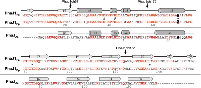
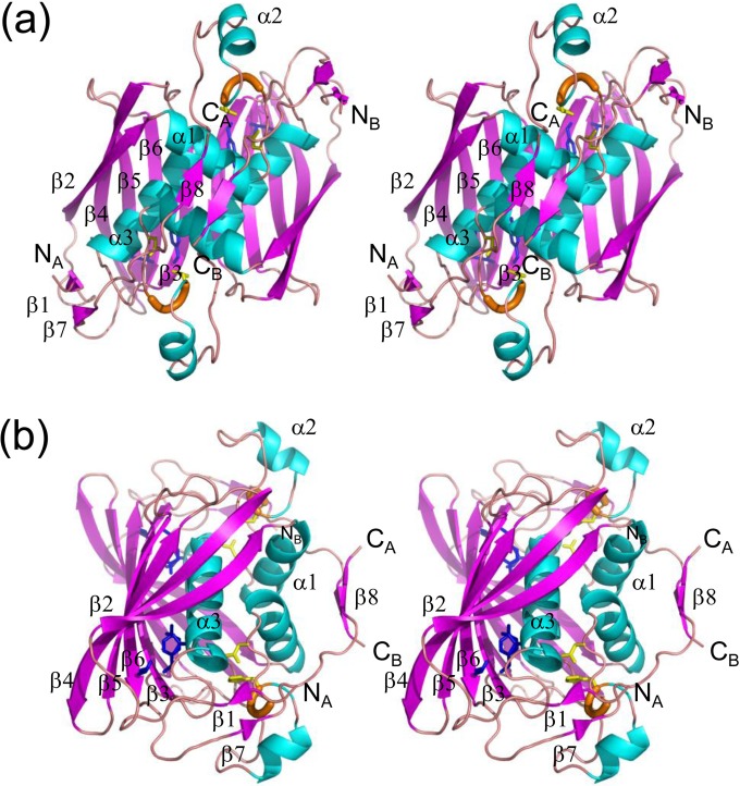
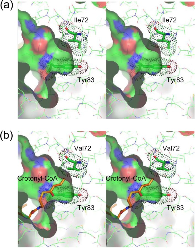
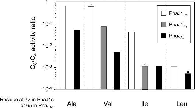
(R)-Specific enoyl-coenzyme A (enoyl-CoA) hydratases (PhaJs) are capable of supplying monomers from fatty acid β-oxidation to polyhydroxyalkanoate (PHA) biosynthesis. PhaJ1Pp from Pseudomonas putida showed broader substrate specificity than did PhaJ1Pa from Pseudomonas aeruginosa, despite sharing 67% amino acid sequence identity. In this study, the substrate specificity characteristics of two Pseudomonas PhaJ1 enzymes were investigated by site-directed mutagenesis, chimeragenesis, X-ray crystallographic analysis, and homology modeling. In PhaJ1Pp, the replacement of valine with isoleucine at position 72 resulted in an increased preference for enoyl-coenzyme A (CoA) elements with shorter chain lengths. Conversely, at the same position in PhaJ1Pa, the replacement of isoleucine with valine resulted in an increased preference for enoyl-CoAs with longer chain lengths. These changes suggest a narrowing and broadening in the substrate specificity range of the PhaJ1Pp and PhaJ1Pa mutants, respectively. However, the substrate specificity remains broader in PhaJ1Pp than in PhaJ1Pa. Additionally, three chimeric PhaJ1 enzymes, composed from PhaJ1Pp and PhaJ1Pa, all showed significant hydratase activity, and their substrate preferences were within the range exhibited by the parental PhaJ1 enzymes. The crystal structure of PhaJ1Pa was determined at a resolution of 1.7 Å, and subsequent homology modeling of PhaJ1Pp revealed that in the acyl-chain binding pocket, the amino acid at position 72 was the only difference between the two structures. These results indicate that the chain-length specificity of PhaJ1 is determined mainly by the bulkiness of the amino acid residue at position 72, but that other factors, such as structural fluctuations, also affect specificity.


Figures

FIG 1
FIG 1
Amino acid sequence alignment of PhaJ1Pa from P. aeruginosa DSM1707 and PhaJ1Pp from P. putida KT2440, with PhaJAc from A. caviae. Secondary structures of PhaJ1Pa and PhaJ1Ac are indicated along with each of their sequences. Helices and strands are shown by cylinders and arrows, respectively. Conserved residues in PhaJ1Pa and PhaJ1Pp are shown in red, whereas those in all three enzymes are indicated in boldface. Active-site residues Asp38 and His43 are indicated by the number sign, and residues Ile/Val72, Tyr83, and Ile136, which constitute the acyl-chain-binding pocket in PhaJ1, are indicated by asterisks. Vertical arrows indicate the junction points of the chimeric PhaJ1 enzymes (P. aeruginosa PhaJ1 is the N terminus of the chimera, and P. putida PhaJ1 is the C terminus).
FIG 2
FIG 2
Stereo diagrams of the crystal structure of the PhaJ1Pa dimer. α-Helices, β-strands, and 310-helices are indicated by helices (blue), arrows (magenta), and thick tubes (orange), respectively. The catalytic dyad residues Asp38 and His43 are represented by stick models (yellow). Residues located at the bottom of the acyl-chain-binding pocket (Ile72, Tyr83, and Ile136) also are represented by stick models (blue). N and C termini of each chain are labeled. Secondary structure annotations are shown only for chain A. (a) A front view of the structure along the 2-fold axis of symmetry. Two subunits adopt an almost identical conformation, with an RMSD of 0.38 Å for Cα atoms. (b) A side view of the structure, generated by 90° rotation relative to the image shown in panel a, highlighting the four-layered structure.
FIG 3
FIG 3
χ1-χ2 plot for eight tyrosine residues in PhaJ1Pa generated by PROCHECK (33). Commonly observed side-chain conformations for tyrosine residues are shown as green regions. Tyr83 residues in chains A and B of PhaJ1Pa are indicated by red boxes. The other six residues are indicated by yellow boxes.
FIG 4
FIG 4
Stereo diagrams of the molecular surfaces of the acyl-chain binding pockets of PhaJ1Pa (a) and PhaJ1Pp (b). Molecular surfaces, calculated with a probe radius of 1.7 Å, are represented by a solid surface using a cutaway view. Residues are represented by thin-stick models with carbon, oxygen, nitrogen, and sulfur atoms in green, red, blue, and yellow, respectively. Residues Ile72 and Tyr83 are represented by thick-stick models. van der Waals surfaces of these residues are shown as dots. (b) Crotonyl-CoA, represented by an orange stick model, is manually docked in the substrate-binding pocket of PhaJ1Pp by using the binding mode of (R)-3-hydroxydecanoyl-CoA bound to fungal hydratase 2 (PDB entry 1PN4) (12) as a reference model.
FIG 5
FIG 5
Comparison of C8/C4 activity ratios. Activity ratios are represented as white bars for PhaJ1Pp and its mutants, gray bars for PhaJ1Pa and its mutant, and black bars for PhaJAc and its mutants. Wild-type enzymes are indicated by an asterisk. Data for PhaJAc and its mutants were retrieved from reference .

References

    1. Madison LL, Huisman GW. 1999. Metabolic engineering of poly(3-hydroxyalkanoates): from DNA to plastic. Microbiol Mol Biol Rev 63:21–53. - PMC - PubMed
    1. Lenz RW, Marchessault RH. 2005. Bacterial polyesters: biosynthesis, biodegradable plastics and biotechnology. Biomacromolecules 6:1–8. doi:10.1021/bm049700c. - DOI - PubMed
    1. Sudesh K, Abe H, Doi Y. 2000. Synthesis, structure and properties of polyhydroxyalkanoate: biological polyesters. Prog Polym Sci 25:1503–1555. doi:10.1016/S0079-6700(00)00035-6. - DOI
    1. Fukui T, Doi Y. 1997. Cloning and analysis of the poly(3-hydroxybutyrate-co-3-hydroxyhexanoate) biosynthesis genes of Aeromonas caviae. J Bacteriol 179:4821–4830. - PMC - PubMed
    1. Fukui T, Shiomi N, Doi Y. 1998. Expression and characterization of (R)-specific enoyl coenzyme A hydratase involved in polyhydroxyalkanoate biosynthesis by Aeromonas caviae. J Bacteriol 180:667–673. - PMC - PubMed

MeSH terms

Substances

LinkOut - more resources