. 2015 Dec;81(23):8192-201.
doi: 10.1128/AEM.02622-15. Epub 2015 Sep 18.

Poultry body temperature contributes to invasion control through reduced expression of Salmonella pathogenicity island 1 genes in Salmonella enterica serovars Typhimurium and Enteritidis

Affiliations

Poultry body temperature contributes to invasion control through reduced expression of Salmonella pathogenicity island 1 genes in Salmonella enterica serovars Typhimurium and Enteritidis

Bryan Troxell et al. Appl Environ Microbiol. 2015 Dec.

Abstract

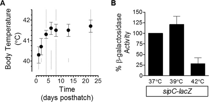
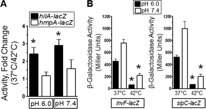
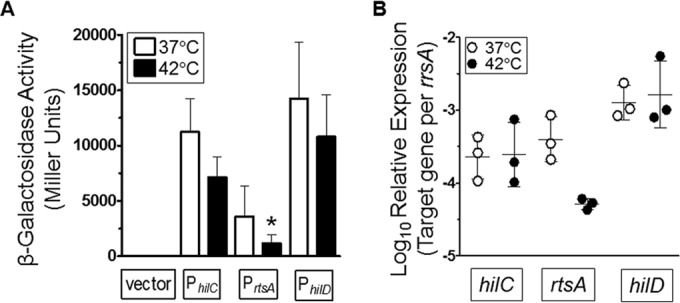
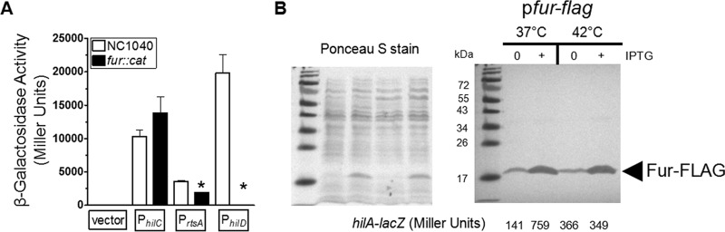
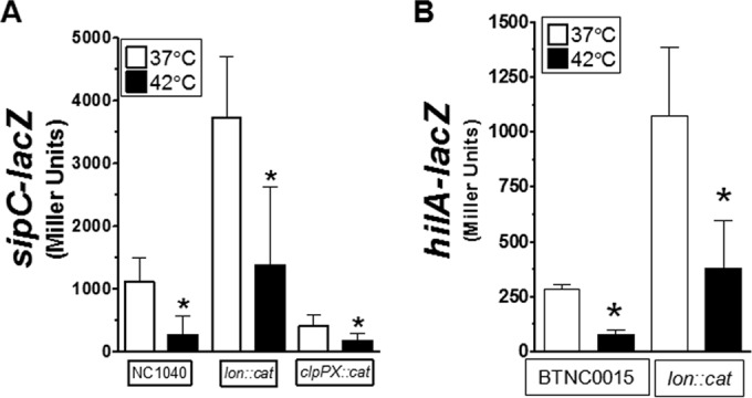
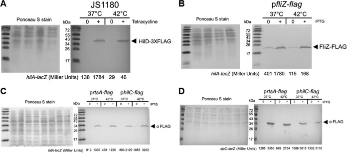
Salmonella enterica serovars Typhimurium (S. Typhimurium) and Enteritidis (S. Enteritidis) are foodborne pathogens, and outbreaks are often associated with poultry products. Chickens are typically asymptomatic when colonized by these serovars; however, the factors contributing to this observation are uncharacterized. Whereas symptomatic mammals have a body temperature between 37°C and 39°C, chickens have a body temperature of 41°C to 42°C. Here, in vivo experiments using chicks demonstrated that numbers of viable S. Typhimurium or S. Enteritidis bacteria within the liver and spleen organ sites were ≥4 orders of magnitude lower than those within the ceca. When similar doses of S. Typhimurium or S. Enteritidis were given to C3H/HeN mice, the ratio of the intestinal concentration to the liver/spleen concentration was 1:1. In the avian host, this suggested poor survival within these tissues or a reduced capacity to traverse the host epithelial layer and reach liver/spleen sites or both. Salmonella pathogenicity island 1 (SPI-1) promotes localization to liver/spleen tissues through invasion of the epithelial cell layer. Following in vitro growth at 42°C, SPI-1 genes sipC, invF, and hilA and the SPI-1 rtsA activator were downregulated compared to expression at 37°C. Overexpression of the hilA activators fur, fliZ, and hilD was capable of inducing hilA-lacZ at 37°C but not at 42°C despite the presence of similar levels of protein at the two temperatures. In contrast, overexpression of either hilC or rtsA was capable of inducing hilA and sipC at 42°C. These data indicate that physiological parameters of the poultry host, such as body temperature, have a role in modulating expression of virulence.


Figures

FIG 1
FIG 1
Expression of the SPI-1 gene sipC is repressed following growth at 42°C. (A) The body temperatures of a W-36 line of chickens were measured 1, 2, 4, 6, 8, 13, and 22 days posthatch (indicated by dashed lines). By day 8, the body temperature was not significantly different than at day 6 (P < 0.05 [Student's t test and Bonferroni adjustment for multiple comparisons]). (B) A sipC-lacZ fusion strain (RM5385) was used to test the impact of different temperatures on the regulation of SPI-1. Bacteria were grown overnight in LB medium under standing conditions at 37°C and diluted 50-fold in LB medium containing MOPS (pH 7.4, 100 mM) and glucose (1 mM) and incubated at different temperatures under standing conditions for approximately 16 to 18 h. β-Galactosidase activity was determined from experiments performed on three separate occasions. Data shown are the means ± SD, with reporter activity under the 37°C conditions set to 100%.
FIG 2
FIG 2
Growth at 42°C repressed SPI-1 genes independently of pH. (A) Bacteria were grown as described in the Fig. 1B legend. The pH of the medium was left unchanged or buffered to 7.4 with NaOH. Bacteria were grown overnight, and β-galactosidase activity was determined. The hmpA-lacZ strain was used as a control. Data are from 4 separate experiments, and an asterisk indicates a significant difference in the fold change measured for the hilA-lacZ strain compared to the control, the hmpA-lacZ strain. Strains used were RM5948 and AV0305. (B) Bacteria were treated as described for panel A, and the reporter activity of strains was measured. Data are from 3 separate experiments, and an asterisk indicates a significant difference in reporter activity at 42°C compared to 37°C under the same pH conditions. Strains used were CA701 and RM5385.
FIG 3
FIG 3
The promoters of the SPI-1 activators, hilC and rtsA, are differentially regulated by growth at 42°C. (A) The promoters of hilC, rtsA, and hilD were cloned into the pSP417 multicopy shuttle vector (empty vector) upstream of a promoterless lacZ gene. Bacteria were grown as described for Fig. 1B and diluted 1:50 into LB-MOPS medium with 1 mM glucose at pH 7.4 at 37°C or 42°C. β-Galactosidase activity was measured after overnight growth. Data are from 4 separate experiments, and a statistically significant result compared to activity at 37°C was determined. The strains used were BTNC0002 to BTNC0005. (B) Bacteria were grown in LB-MOPS medium with 1 mM glucose at pH 7.4 at 37°C or 42°C. Total RNA was extracted at an OD600 of ∼1. cDNA was generated using gene-specific primers, and expression data were normalized to the rrsA 16S rRNA gene.
FIG 4
FIG 4
Growth at 42°C repressed expression of SPI-1 genes independently of the activator Fur. (A) The promoters of hilC, rtsA, and hilD were cloned into the pSP417 multicopy shuttle vector (empty vector) upstream of a promoterless lacZ gene. Bacteria were grown as described for Fig. 1B, and the promoter activities were determined for the NC1040 and fur::cat strains following overnight growth at 37°C. Data are from 3 separate experiments. The strains used were BTNC0002 to BTNC0005 and BTNC0006 to BTNC0009. (B) Expression of a hilA-lacZ fusion at the chromosomal att site was determined following overnight growth with or without induction of the Fur-FLAG protein. Bacteria were grown as described for Fig. 1B, except that samples were diluted 500-fold and cultivated at either 37°C or 42°C. A portion of each sample was removed to measure β-galactosidase activity, and the remainder was treated for SDS-PAGE and Western blotting to detect the FLAG epitope. Following transfer of proteins to the nitrocellulose membrane, the membrane was stained with Ponceau S to ensure that equivalent levels of protein were loaded for samples and that ∼2 × 10⁸ cells were loaded per lane (left panel). IPTG was added to the growth medium to reach a concentration of 0.1 mM to induce Fur-FLAG. Western blotting with the anti-FLAG antibody revealed cross-reactivity to the Fur protein of the expected size, indicated by the arrowhead with the appropriate label (right panel). The β-galactosidase activity for each sample is listed below each lane (right panel). Samples shown are representative of the results of 3 separate experiments. The complete β-galactosidase activity data are listed here. For BTNC0017 (pfur-flag), the values measured at 37°C were 156 ± 33 under uninduced conditions and 657 ± 89 under induced conditions and the values measured at 42°C were 316 ± 70 under uninduced conditions and 345 ± 4 under induced conditions.
FIG 5
FIG 5
The Lon heat shock protease contributes to the basal level of SPI-1 repression but does not perturb inactivation following growth at 42°C. (A) The sipC-lacZ fusion was used to determine the contribution of lon or clpPX to the regulation of SPI-1 following growth at 42°C. Bacteria were grown as described for Fig. 1B, and reporter activity was measured after overnight growth. The strains used were BTNC0010 to BTNC0012. (B) The hilA-lacZ fusion integrated into the att within the chromosome was used to determine the influence of lon on the reduced expression following growth at 42°C. Samples were cultivated as described for Fig. 1B after overnight growth. Data are from 4 separate experiments. The strains used were BTNC0015 and BTNC0016.
FIG 6
FIG 6
Overexpression of either the HilD or the FliZ SPI-1 activator does not activate SPI-1 at 42°C, but overexpression of either HilC or RtsA does. (A) Expression of a hilA-lacZ fusion at the chromosomal att site was determined following overnight growth with or without induction of the HilD-3×FLAG protein. Bacteria were grown as described for Fig. 1B, except that samples were diluted 500-fold and cultivated at either 37°C or 42°C. A portion of each sample was removed to measure β-galactosidase activity, and the remainder was treated for SDS-PAGE and Western blotting to detect the FLAG epitope. Following transfer of proteins to the nitrocellulose membrane, the membrane was stained with Ponceau S to ensure the equivalent levels of protein were loaded for samples and that ∼2 × 10⁸ cells were loaded per lane (left panel). Tetracycline was added to reach a concentration of 2.5 μg/ml to induce HilD-3×FLAG. Western blotting with the anti-FLAG antibody revealed cross-reactivity to the HilD protein of the expected size, indicated by the arrowhead with the appropriate label (right panel). The β-galactosidase activity for each sample is listed below each lane (right panel). Samples shown are representative of the results of 3 separate experiments. The complete β-galactosidase activity data are listed here. For JS1180, the values measured at 37°C were 133 ± 13 under uninduced conditions and 1,716 ± 146 under induced conditions and the values measured at 42°C were 28 ± 1 under uninduced conditions and 62 ± 34 under induced conditions. (B) Expression of a hilA-lacZ fusion at the chromosomal att site was determined following overnight growth with or without induction of FliZ-FLAG. Samples were prepared as described for panel A for the Ponceau S staining (left panel), and the Western blot analysis was performed with anti-FLAG (right panel). IPTG was used at 0.1 mM to induce FliZ-FLAG. Sample data shown are representative of the results of 3 separate experiments. The complete β-galactosidase activity data for BTNC0018 (pfliZ-flag) are listed here; the values measured at 37°C were 527 ± 133 under uninduced conditions and 1,539 ± 243 under induced conditions, and the values measured at 42°C were 89 ± 36 under uninduced conditions and 177 ± 92 under induced conditions. (C) Samples were treated as described for panel A for the RtsA-FLAG or HilC-FLAG strains containing the hilA-lacZ fusion and induced with 0.1 mM IPTG. Samples data are representative, and the β-galactosidase activity for each sample is shown below each lane. The complete β-galactosidase activity data are listed here. For BTNC0019 (prtsA-flag), the values measured at 37°C were 580 ± 30 under uninduced conditions and 1,490 ± 317 under induced conditions, and the values measured at 42°C were 367 ± 66 under uninduced conditions and 2,177 ± 504 under induced conditions. For BTNC0020 (philC-flag), the values measured at 37°C were 811 ± 9 under uninduced conditions and 1,936 ± 319 under induced conditions, and the values measured at 42°C were 965 ± 113 under uninduced conditions and 3,142 ± 649 under induced conditions. (D) Strain RM5385 was transformed with prtsA-flag or philC-flag and reporter activity was determined at different temperatures. IPTG was used at 0.01 mM to induce RtsA-FLAG and HilC-FLAG. The Ponceau S (left panel) and Western blotting (right panel) are shown. The complete β-galactosidase activity data are listed here. For BTNC0023 (prtsA-flag), the values measured at 37°C were 1,966 ± 781 under uninduced conditions and 3,409 ± 103 under induced conditions, and the values measured at 42°C were 712 ± 469 under uninduced conditions and 2,666 ± 171 under induced conditions. For BTNC0024 (philC-flag), the values measured at 37°C were 2,801 ± 769 under uninduced conditions and 3,727 ± 906 under induced conditions, and the values measured at 42°C were 1,774 ± 521 under uninduced conditions and 3,122 ± 790 under induced conditions.

Similar articles

Cited by

References

    1. CDC. 2011. National Salmonella surveillance annual report. US Department of Health and Human Services, CDC, Atlanta, GA.
    1. CDC. 2013. Surveillance for foodborne disease outbreaks—United States, 2009–2010. MMWR Morb Mortal Wkly Rep 62:41–47. - PMC - PubMed
    1. Ellermeier JR, Slauch JM. 2007. Adaptation to the host environment: regulation of the SPI1 type III secretion system in Salmonella enterica serovar Typhimurium. Curr Opin Microbiol 10:24–29. doi:10.1016/j.mib.2006.12.002. - DOI - PubMed
    1. Eichelberg K, Galan JE. 1999. Differential regulation of Salmonella typhimurium type III secreted proteins by pathogenicity island 1 (SPI-1)-encoded transcriptional activators InvF and hilA. Infect Immun 67:4099–4105. - PMC - PubMed
    1. Altier C. 2005. Genetic and environmental control of salmonella invasion. J Microbiol 43(Spec No):85–92. - PubMed

Publication types

MeSH terms

LinkOut - more resources