Review
. 2015 Nov;309(9):H1453-67.
doi: 10.1152/ajpheart.00554.2015. Epub 2015 Sep 18.

Drug-induced mitochondrial dysfunction and cardiotoxicity

Affiliations
Review

Drug-induced mitochondrial dysfunction and cardiotoxicity

Zoltán V Varga et al. Am J Physiol Heart Circ Physiol. 2015 Nov.

Abstract

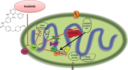
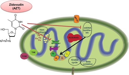
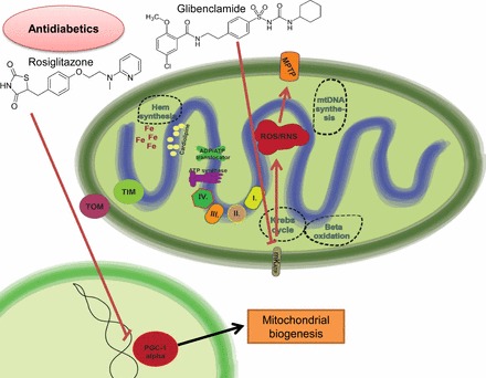
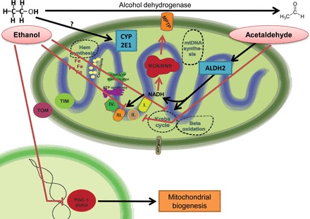
Mitochondria has an essential role in myocardial tissue homeostasis; thus deterioration in mitochondrial function eventually leads to cardiomyocyte and endothelial cell death and consequent cardiovascular dysfunction. Several chemical compounds and drugs have been known to directly or indirectly modulate cardiac mitochondrial function, which can account both for the toxicological and pharmacological properties of these substances. In many cases, toxicity problems appear only in the presence of additional cardiovascular disease conditions or develop months/years following the exposure, making the diagnosis difficult. Cardiotoxic agents affecting mitochondria include several widely used anticancer drugs [anthracyclines (Doxorubicin/Adriamycin), cisplatin, trastuzumab (Herceptin), arsenic trioxide (Trisenox), mitoxantrone (Novantrone), imatinib (Gleevec), bevacizumab (Avastin), sunitinib (Sutent), and sorafenib (Nevaxar)], antiviral compound azidothymidine (AZT, Zidovudine) and several oral antidiabetics [e.g., rosiglitazone (Avandia)]. Illicit drugs such as alcohol, cocaine, methamphetamine, ecstasy, and synthetic cannabinoids (spice, K2) may also induce mitochondria-related cardiotoxicity. Mitochondrial toxicity develops due to various mechanisms involving interference with the mitochondrial respiratory chain (e.g., uncoupling) or inhibition of the important mitochondrial enzymes (oxidative phosphorylation, Szent-Györgyi-Krebs cycle, mitochondrial DNA replication, ADP/ATP translocator). The final phase of mitochondrial dysfunction induces loss of mitochondrial membrane potential and an increase in mitochondrial oxidative/nitrative stress, eventually culminating into cell death. This review aims to discuss the mechanisms of mitochondrion-mediated cardiotoxicity of commonly used drugs and some potential cardioprotective strategies to prevent these toxicities.

Keywords: cardiomyopathy; drug development; heart; heart failure; reactive oxygen species; toxicology.


Figures

Fig. 1.
Fig. 1.
Doxorubicin-induced mitochondrial dysfunction and the effect of trastuzumab on mitochondria-related survival pathways. Doxorubicin leads to marked induction of mitochondrial reactive oxygen species (ROS) production. It shows specific binding activity to the mitochondrial abundant cardiolipin, leading to selective mitochondrial accumulation. Doxorubicin is prone to redox cycling, thereby promoting ROS and reactive nitrogen species (RNS) production. There is also increased intramitochondrial free iron accumulation after doxorubicin exposition, giving rise to additional nonenzymatic ROS production by the Haber-Weiss reaction. The major consequences of uncontrolled ROS/RNS production are mitochondrial permeability transition pore (MPTP) opening and poly(ADP-ribose) polymerase (PARP) activation, converging to the propagation to cell death signaling mechanisms. In parallel with ROS/RNS induction, doxorubicin also interferes with the action of topoisomerase-2β, being involved in the regulation of peroxisome proliferator-activated receptor-γ coactivator-1α (PGC-1α) and thereby mitochondrial biogenesis- and metabolism-related pathways. Altogether, these doxorubicin-induced alterations profoundly alter both mitochondrial structure and function. The release of neuregulin-1 by the coronary endothelium activates human epidermal growth factor receptor (Her)4 (ErbB4) to dimerize with Her2 (ErbB2). The Her4/Her2 (ErbB4/ErbB2) dimer activates cardioprotective signaling pathways, including phosphatidyinositol 3-kinase (PI3K)/protein kinase B (Akt), ERK1/2, and focal adhesion kinase (FAK), which promote cell survival upon cellular stress. Trastuzumab blocks Her2 signaling and disrupts this cardioprotective machinery, resulting in loss of cytoprotective mechanisms. Furthermore, trastuzumab may trigger cellular oxidative stress and induce the expression and activation of proapoptotic proteins [e.g., Bcl-2-associated X protein (BAX)]. These events result in mitochondrial defects, leading to the opening of the MPTP and the activation of cell death pathways that precipitate myocardial dysfunction. BAD, Bcl-2-associated death promoter; mKATP, mitochondrial ATP-sensitive potassium channel.
Fig. 2.
Fig. 2.
Mechanisms of imatinib cardiotoxicity. The small molecule inhibitor of the Bcr/Abl (fusion kinase of the break point cluster region of chromosome 22 and the Abelson1 gene of chromosome 9) kinase imatinib induces mitochondrial dysfunction by interfering with mitochondrial protein import machinery. Increased PRKR-like endoplasmic reticulum kinase (PERK) activation in the endoplasmic reticulum leads to the phosphorylation eukaryotic initiation factor 2α (eIF2α) and to translational attenuation of translocase of the inner membrane (TIM) 23-kDa form that is essential for protein import into the mitochondrial matrix. Impaired protein import will negatively affect major mitochondrial metabolic pathways, including mitochondrial DNA synthesis, the Krebs cycle, β-oxidation, and hem synthesis. TOM, translocase of the outer membrane.
Fig. 3.
Fig. 3.
Mechanisms of zidovudin [azidothymidine (AZT)] cardiotoxicity. AZT interferes with mitochondrial DNA polymerase-γ, the enzyme responsible for mtDNA replication. Since the mitochondrial DNA (mtDNA) codes the proteins of the electron transport chain (ETC), inhibition of mtDNA replication will result in mitochondrial energetic imbalance. AZT may directly inhibit the mitochondrial ADP/ATP tranlocator, inhibiting the transport of ATP produced from oxidative phosphorylation and the substrate ADP to be transported from the cytoplasm to the mitochondrial matrix.
Fig. 4.
Fig. 4.
Mechanisms of antidiabetic-related cardiotoxicity. The insulin sensitizer “glitazone” may promote the degradation of PGC-1α, a master regulator of mitochondrial biogenesis and metabolism. Sulfanylureas (e.g., glibenclamide) directly inhibit the mKATPs that are involved in cardioprotective mechanisms. Inhibition of the mKATP leads to increased ROS production and MPTP opening.
Fig. 5.
Fig. 5.
Mechanisms of ethanol cardiotoxicity. Ethanol will increase mitochondrial ROS production by complex mechanisms. On the one hand, ethanol and acetaldehyde will inhibit the function of the Krebs cycle and the ETC, whereas in parallel there is increased intramitochondrial NADH production by aldehyde dehydrogenase 2 (ALDH2). To bypass the inhibited ETC, NADH may be used for ROS/RNS production, leading to MPTP opening. Inhibition of PGC-1α by ethanol leads to deteriorated mitochondrial biogenesis and oxidative metabolism. CYP2E1, cytochrome P450 2E1.

References

    1. Arcamone F, Cassinelli G, Fantini G, Grein A, Orezzi P, Pol C, Spalla C. Adriamycin, 14-hydroxydaunomycin, a new antitumor antibiotic from S. peucetius var. caesius. Biotechnol Bioeng 11: 1101–1110, 1969. - PubMed
    1. Archer SL. Mitochondrial dynamics—mitochondrial fission and fusion in human diseases. N Engl J Med 369: 2236–2251, 2013. - PubMed
    1. Armstrong GT, Joshi VM, Ness KK, Marwick TH, Zhang N, Srivastava D, Griffin BP, Grimm RA, Thomas J, Phelan D, Collier P, Krull KR, Mulrooney DA, Green DM, Hudson MM, Robison LL, Plana JC. Comprehensive Echocardiographic Detection of Treatment-Related Cardiac Dysfunction in Adult Survivors of Childhood Cancer: Results From the St. Jude Lifetime Cohort Study. J Am Coll Cardiol 65: 2511–2522, 2015. - PMC - PubMed
    1. Awtry EH, Philippides GJ. Alcoholic and cocaine-associated cardiomyopathies. Prog Cardiovasc Dis 52: 289–299, 2010. - PubMed
    1. Bai P, Mabley JG, Liaudet L, Virag L, Szabo C, Pacher P. Matrix metalloproteinase activation is an early event in doxorubicin-induced cardiotoxicity. Oncol Rep 11: 505–508, 2004. - PubMed

Publication types

MeSH terms