. 2015 Dec;160(6):1296-1308.e2.
doi: 10.1016/j.ajo.2015.09.015. Epub 2015 Sep 18.

Thinner Retinal Nerve Fiber Layer in Very Preterm Versus Term Infants and Relationship to Brain Anatomy and Neurodevelopment

Affiliations

Thinner Retinal Nerve Fiber Layer in Very Preterm Versus Term Infants and Relationship to Brain Anatomy and Neurodevelopment

Adam L Rothman et al. Am J Ophthalmol. 2015 Dec.

Abstract

Purpose: To assess retinal nerve fiber layer (RNFL) thickness at term-equivalent age in very preterm (<32 weeks gestational age) vs term-born infant cohorts, and compare very preterm infant RNFL thickness with brain anatomy and neurodevelopment.

Design: Cohort study.

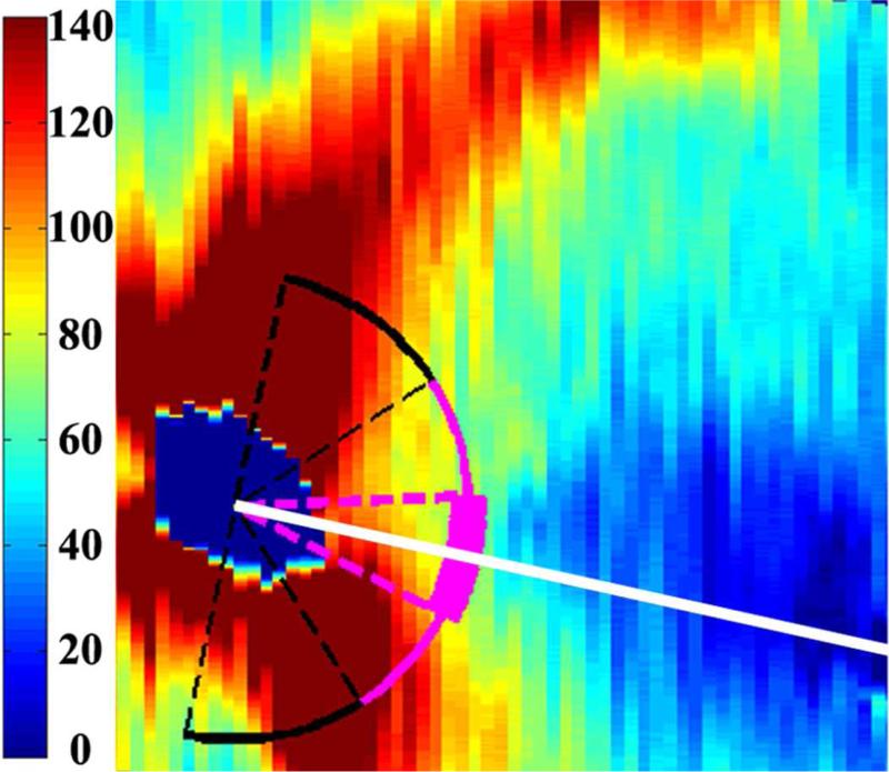
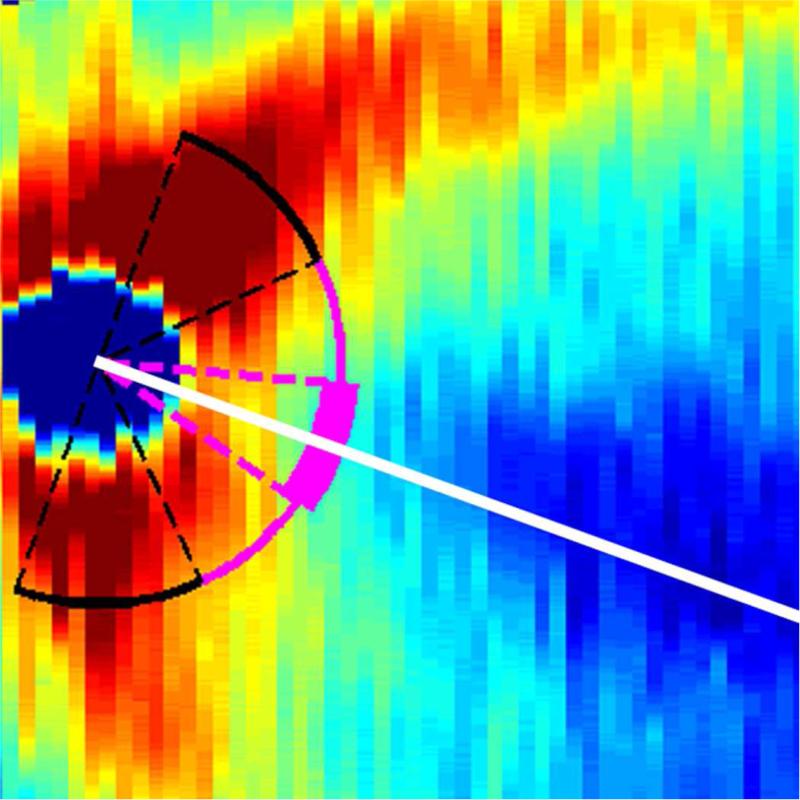
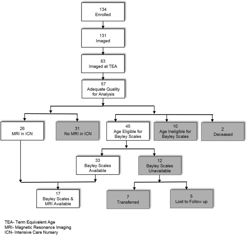
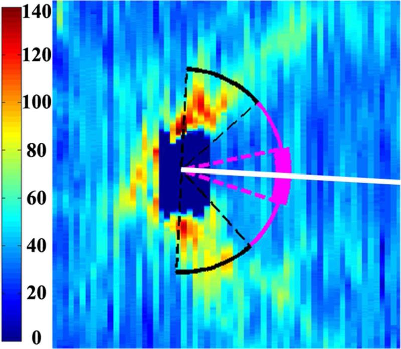
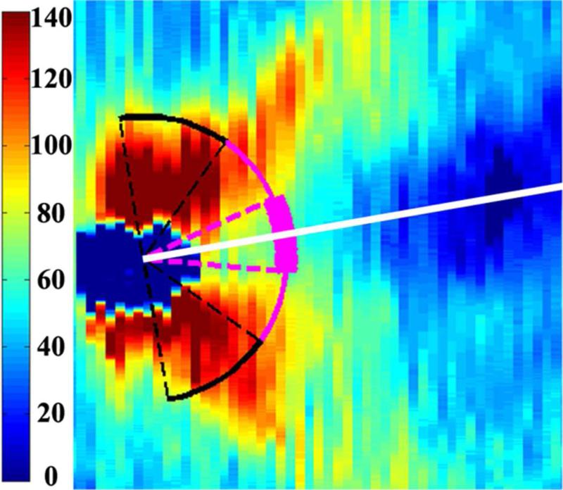
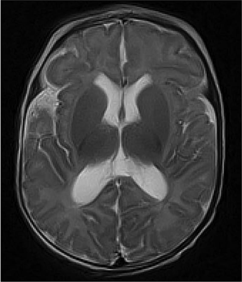
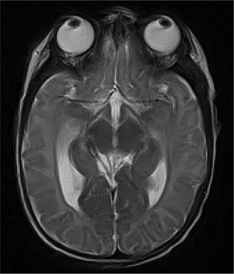
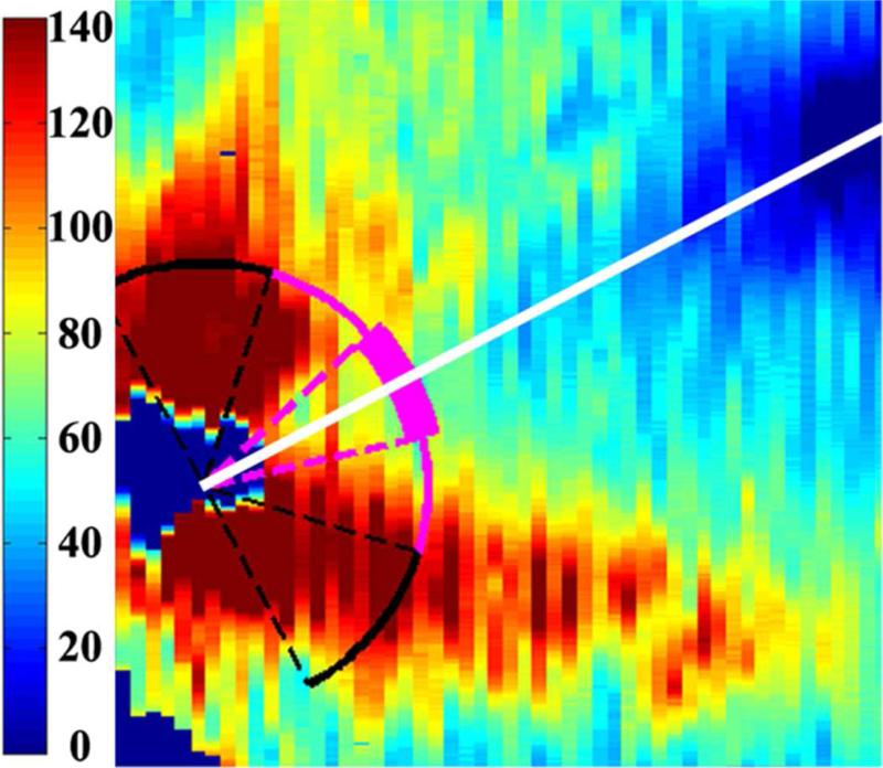
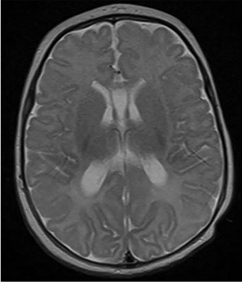
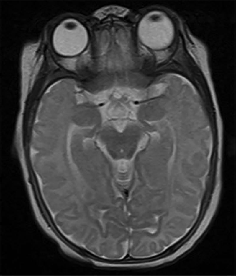
Methods: RNFL was semi-automatically segmented (1 eye per infant) in 57 very preterm and 50 term infants with adequate images from bedside portable, handheld spectral-domain optical coherence tomography imaging at 37-42 weeks postmenstrual age. Mean RNFL thickness was calculated for the papillomacular bundle (-15 degrees to +15 degrees) and temporal quadrant (-45 degrees to +45 degrees) relative to the fovea-optic nerve axis. Brain magnetic resonance imaging (MRI) scans clinically obtained in 26 very preterm infants were scored for global structural abnormalities by an expert masked to data except for age. Cognitive, language, and motor skills were assessed in 33 of the very preterm infants at 18-24 months corrected age.

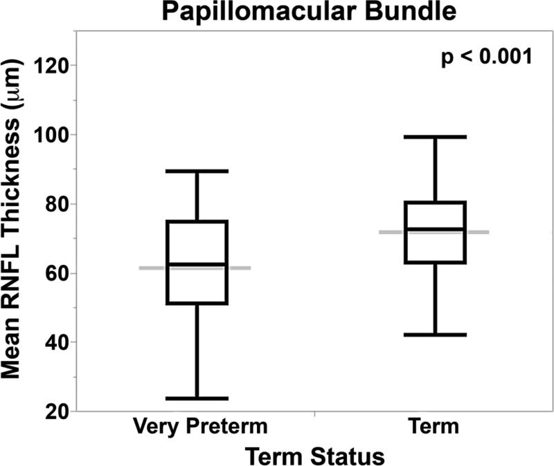
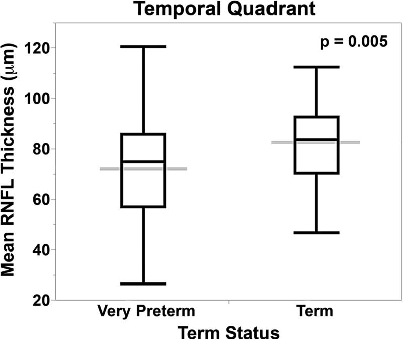
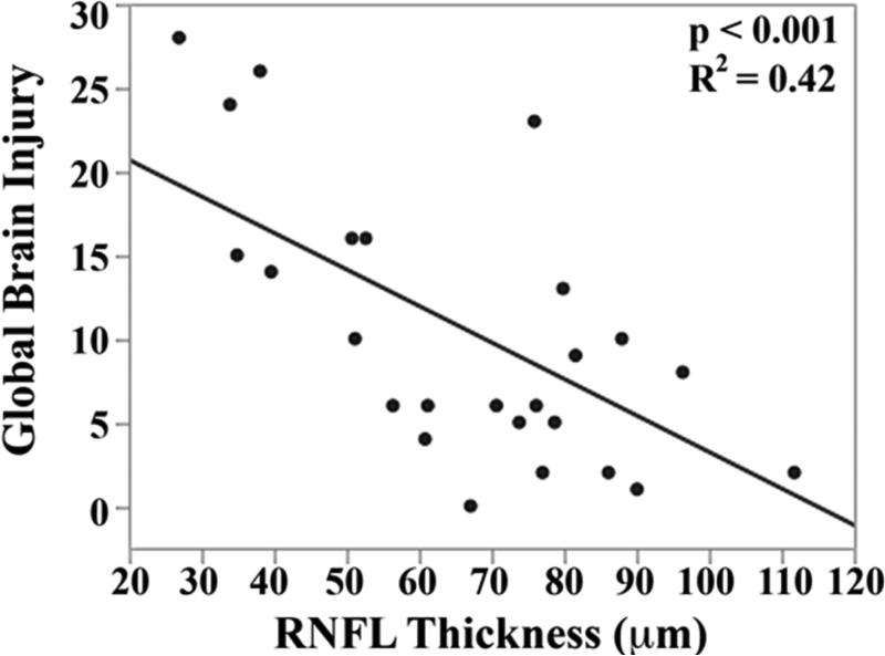
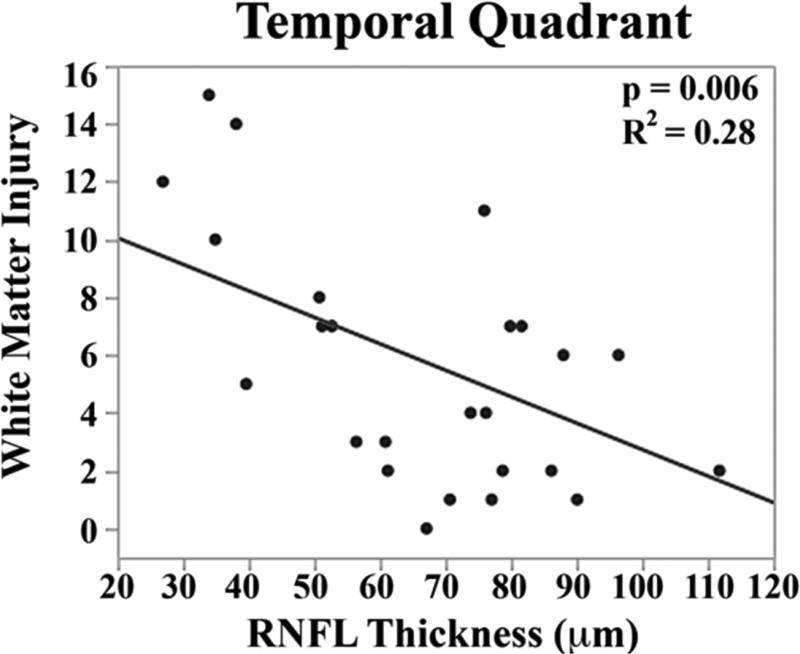
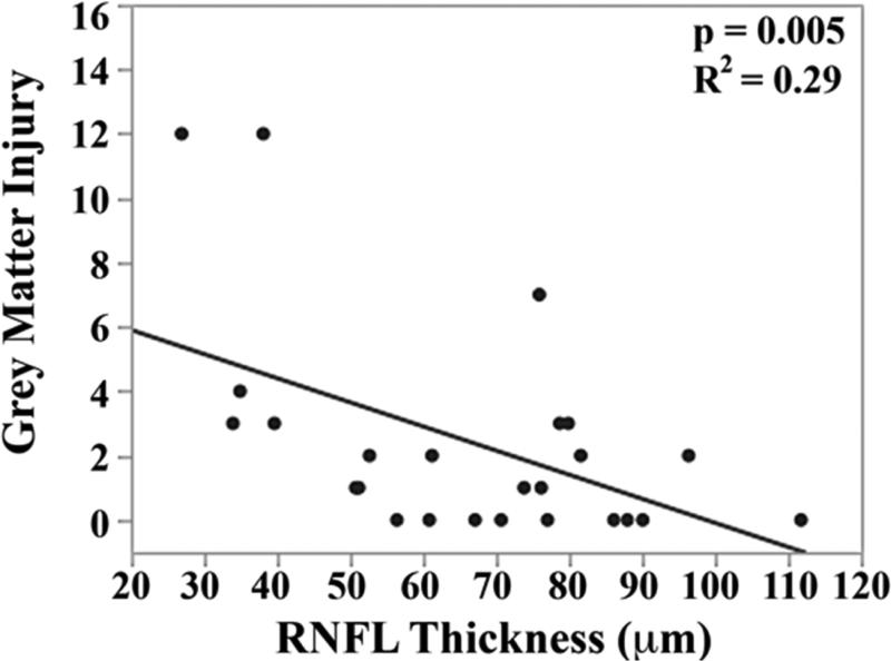
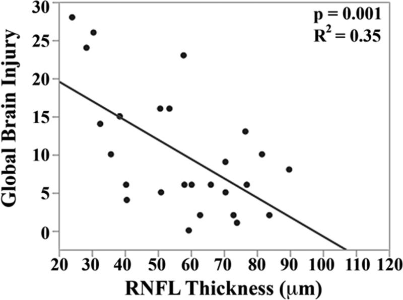
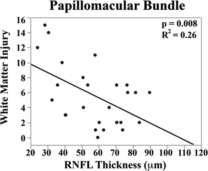
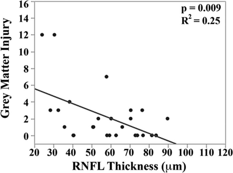
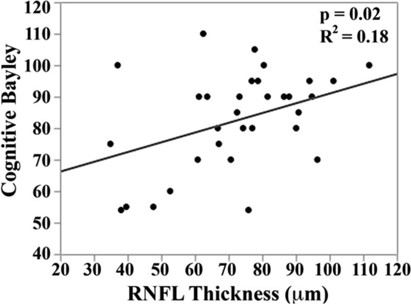
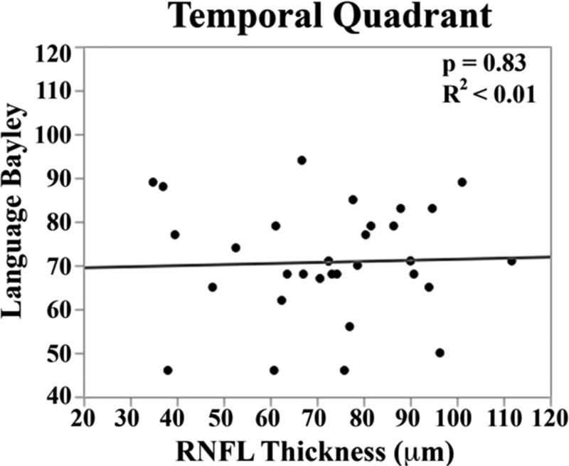
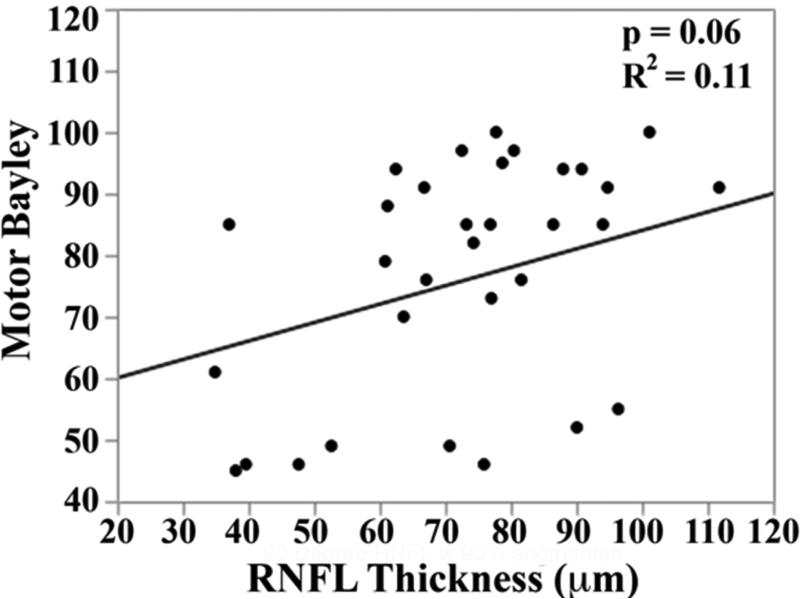
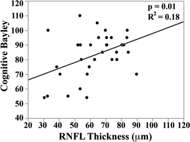
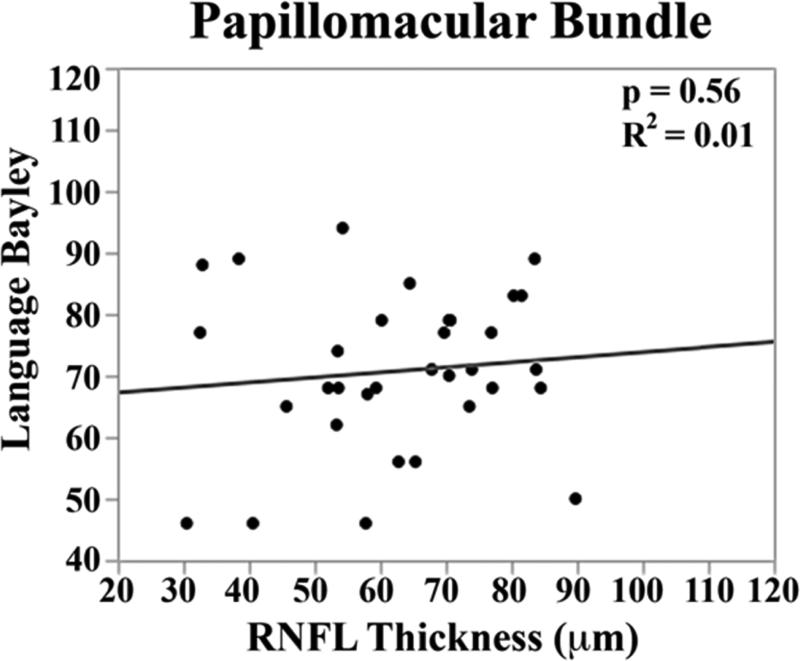
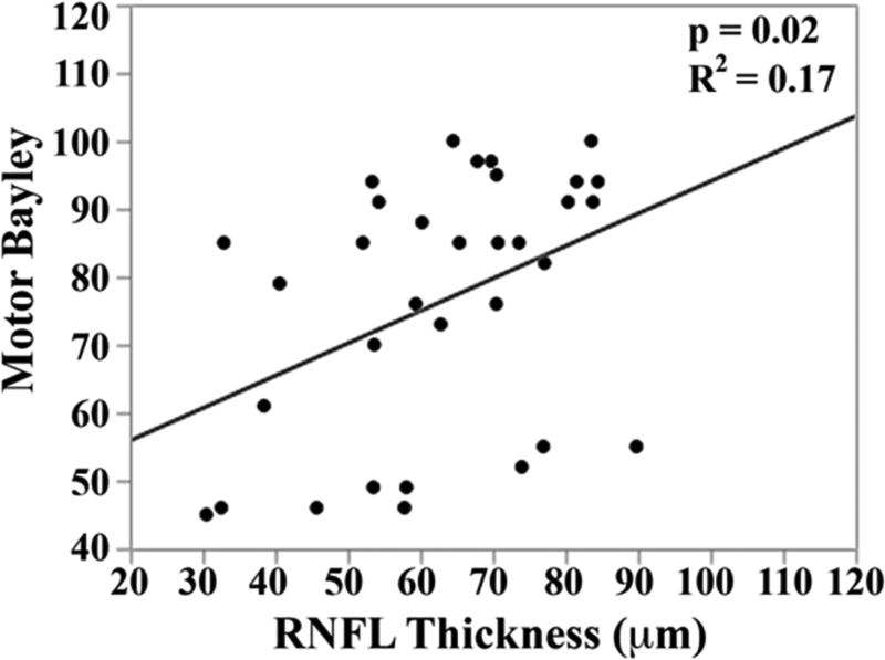
Results: RNFL was thinner for very preterm vs term infants at the papillomacular bundle ([mean ± standard deviation] 61 ± 17 vs 72 ± 13 μm, P < .001) and temporal quadrant (72 ± 21 vs 82 ± 16 μm, P = .005). In very preterm infants, thinner papillomacular bundle RNFL correlated with higher global brain MRI lesion burden index (R(2) = 0.35, P = .001) and lower cognitive (R(2) = 0.18, P = .01) and motor (R(2) = 0.17, P = .02) scores. Relationships were similar for temporal quadrant.

Conclusions: Thinner RNFL in very preterm infants relative to term-born infants may relate to brain structure and neurodevelopment.


Figures

Figure 1
Figure 1
Retinal nerve fiber layer (RNFL) thickness maps derived from segmented spectral domain optical coherence tomography scans obtained in the nursery. The thick pink arc corresponds to the papillomacular bundle: arc from −15 to +15 degrees relative to the organizing axis from the optic nerve center to the fovea. The thin pink arc corresponds to the temporal quadrant: arc from −45 to +45 degrees relative to the organizing axis. Left, RNFL thickness map of a healthy, term Hispanic male born and imaged at 39 weeks post-menstrual age. Mean RNFL thickness is 83 μm across the papillomacular bundle and 97 μm across the temporal quadrant. Right, RNFL thickness map of a white male born at 31 weeks gestational age and 1400 g with stage 0 retinopathy of prematurity. Mean RNFL thickness at 39 weeks post-menstrual age is 64 μm across the papillomacular bundle and 78 μm across the temporal quadrant.
Figure 1
Figure 1
Retinal nerve fiber layer (RNFL) thickness maps derived from segmented spectral domain optical coherence tomography scans obtained in the nursery. The thick pink arc corresponds to the papillomacular bundle: arc from −15 to +15 degrees relative to the organizing axis from the optic nerve center to the fovea. The thin pink arc corresponds to the temporal quadrant: arc from −45 to +45 degrees relative to the organizing axis. Left, RNFL thickness map of a healthy, term Hispanic male born and imaged at 39 weeks post-menstrual age. Mean RNFL thickness is 83 μm across the papillomacular bundle and 97 μm across the temporal quadrant. Right, RNFL thickness map of a white male born at 31 weeks gestational age and 1400 g with stage 0 retinopathy of prematurity. Mean RNFL thickness at 39 weeks post-menstrual age is 64 μm across the papillomacular bundle and 78 μm across the temporal quadrant.
Figure 2
Figure 2
Infant enrollment and eligibility for analysis. Note that all infants were referred for Bayley Scales assessment while brain magnetic resonance imaging was obtained at the discretion of the neonatology team. As a result, very preterm infants may be included in neither, one, or both secondary analyses.
Figure 3
Figure 3
Mean retinal nerve fiber layer thickness across both the papillomacular bundle and temporal quadrant is thinner in very preterm versus term infants at term-equivalent age. The box and whisker plots represent the median, quartiles, maximums and minimums of each Bayley score for each group. The grey line represents the mean for each group.
Figure 3
Figure 3
Mean retinal nerve fiber layer thickness across both the papillomacular bundle and temporal quadrant is thinner in very preterm versus term infants at term-equivalent age. The box and whisker plots represent the median, quartiles, maximums and minimums of each Bayley score for each group. The grey line represents the mean for each group.
Figure 4
Figure 4
Correlation between mean retinal nerve fiber layer (RNFL) thickness along the papillomacular bundle, top) and temporal quadrant (bottom) and global brain magnetic resonance imaging (MRI) lesion burden index, white matter injury, and grey matter injury for 26 very preterm infants who underwent brain MRI while in the intensive care nursery. Thinner RNFL across either arc (papillomacular bundle or temporal quadrant) correlated with an increase in global brain injury, white matter injury and grey matter injury.
Figure 4
Figure 4
Correlation between mean retinal nerve fiber layer (RNFL) thickness along the papillomacular bundle, top) and temporal quadrant (bottom) and global brain magnetic resonance imaging (MRI) lesion burden index, white matter injury, and grey matter injury for 26 very preterm infants who underwent brain MRI while in the intensive care nursery. Thinner RNFL across either arc (papillomacular bundle or temporal quadrant) correlated with an increase in global brain injury, white matter injury and grey matter injury.
Figure 5
Figure 5
Correlation between mean retinal nerve fiber layer (RNFL) thickness along the papillomacular bundle, top) and temporal quadrant (bottom) and cognitive, language, and motor skills for 33 very preterm infants assessed at 18-24 months corrected age with the Bayley Scales of Infant and Toddler Development, 3rd edition. In very preterm infants, thinner RNFL across the papillomacular bundle weakly correlated with poorer cognitive and motor skills as a toddler and across the temporal quadrant weakly correlated with poorer cognitive skills as a toddler.
Figure 5
Figure 5
Correlation between mean retinal nerve fiber layer (RNFL) thickness along the papillomacular bundle, top) and temporal quadrant (bottom) and cognitive, language, and motor skills for 33 very preterm infants assessed at 18-24 months corrected age with the Bayley Scales of Infant and Toddler Development, 3rd edition. In very preterm infants, thinner RNFL across the papillomacular bundle weakly correlated with poorer cognitive and motor skills as a toddler and across the temporal quadrant weakly correlated with poorer cognitive skills as a toddler.
Figure 6
Figure 6
Very preterm infant retinal nerve fiber layer (RNFL) thickness maps at term equivalent age (left column) and their corresponding near-term T2 weighted brain magnetic resonance imaging (MRI, middle, right columns). Top, Black female born at 24 weeks gestational age and 640 g who developed plus disease requiring laser treatment. RNFL thickness map at 37 weeks post-menstrual age measures mean RNFL thickness across the papillomacular bundle (thick pink arc) and temporal quadrant (thin pink arc) of 73 and 86 μm, respectively. Brain MRI at 44 weeks post-menstrual shows a near normal global brain with global brain MRI lesion burden index of 2 (due to a white matter subscore of 2). Middle, Black female born at 23 weeks gestational age and 475 g who developed stage 3 retinopathy of prematurity requiring laser treatment. RNFL thickness map at 37 weeks post-menstrual age measures mean RNFL thickness across the papillomacular bundleand temporal quadrants of 51 and 74 μm, respectively. Brain MRI at 46 weeks post-menstrual age had a global brain MRI lesion burden index of 5 (white matter subscore of 4 and grey matter score of 1). The ventricles are mildly enlarged, especially in the occipital lobe area, consistent with mild white matter volume loss. Bottom, White male born at 25 weeks gestational age and 810 g who developed stage 3 retinopathy of prematurity requiring laser treatment. Cognitive, language, and motor Bayley subscores at 18-24 months corrected age were 75, 89, and 61, respectively. RNFL thickness map at 38 weeks post-menstrual age had mean RNFL thicknesses across the papillomacular bundle and temporal quadrants of 35 and 38 μm, respectively. Brain MRI at 36 weeks post-menstrual age had a global brain MRI lesion burden index of 15 (white matter subscore of 10 and grey matter subscore of 4). The ventricles are markedly enlarged, consistent with severe white matter volume loss.
Figure 6
Figure 6
Very preterm infant retinal nerve fiber layer (RNFL) thickness maps at term equivalent age (left column) and their corresponding near-term T2 weighted brain magnetic resonance imaging (MRI, middle, right columns). Top, Black female born at 24 weeks gestational age and 640 g who developed plus disease requiring laser treatment. RNFL thickness map at 37 weeks post-menstrual age measures mean RNFL thickness across the papillomacular bundle (thick pink arc) and temporal quadrant (thin pink arc) of 73 and 86 μm, respectively. Brain MRI at 44 weeks post-menstrual shows a near normal global brain with global brain MRI lesion burden index of 2 (due to a white matter subscore of 2). Middle, Black female born at 23 weeks gestational age and 475 g who developed stage 3 retinopathy of prematurity requiring laser treatment. RNFL thickness map at 37 weeks post-menstrual age measures mean RNFL thickness across the papillomacular bundleand temporal quadrants of 51 and 74 μm, respectively. Brain MRI at 46 weeks post-menstrual age had a global brain MRI lesion burden index of 5 (white matter subscore of 4 and grey matter score of 1). The ventricles are mildly enlarged, especially in the occipital lobe area, consistent with mild white matter volume loss. Bottom, White male born at 25 weeks gestational age and 810 g who developed stage 3 retinopathy of prematurity requiring laser treatment. Cognitive, language, and motor Bayley subscores at 18-24 months corrected age were 75, 89, and 61, respectively. RNFL thickness map at 38 weeks post-menstrual age had mean RNFL thicknesses across the papillomacular bundle and temporal quadrants of 35 and 38 μm, respectively. Brain MRI at 36 weeks post-menstrual age had a global brain MRI lesion burden index of 15 (white matter subscore of 10 and grey matter subscore of 4). The ventricles are markedly enlarged, consistent with severe white matter volume loss.

References

    1. Woodward LJ, Clark CA, Bora S, Inder TE. Neonatal white matter abnormalities an important predictor of neurocognitive outcome for very preterm children. PLoS One. 2012;7(12):e51879. - PMC - PubMed
    1. Zhang Y, Inder TE, Neil JJ, et al. Cortical structural abnormalities in very preterm children at 7years of age. Neuroimage. 2015;109:469–479. - PMC - PubMed
    1. Pineda RG, Tjoeng TH, Vavasseur C, Kidokoro H, Neil JJ, Inder T. Patterns of altered neurobehavior in preterm infants within the neonatal intensive care unit. J Pediatr. 2013;162(3):470–476. - PMC - PubMed
    1. Mukherjee P, McKinstry RC. Diffusion tensor imaging and tractography of human brain development. Neuroimaging Clin N Am. 2006;16(1):19–43, vii. - PubMed
    1. Msall ME, Phelps DL, Hardy RJ, et al. Educational and social competencies at 8 years in children with threshold retinopathy of prematurity in the CRYO-ROP multicenter study. Pediatrics. 2004;113(4):790–799. - PubMed

Publication types