. 2015 Dec;27(12):2467-73.
doi: 10.1016/j.cellsig.2015.09.003. Epub 2015 Sep 18.

VCAM-1 is a TGF-β1 inducible gene upregulated in idiopathic pulmonary fibrosis

Affiliations

VCAM-1 is a TGF-β1 inducible gene upregulated in idiopathic pulmonary fibrosis

Marianna Agassandian et al. Cell Signal. 2015 Dec.

Abstract

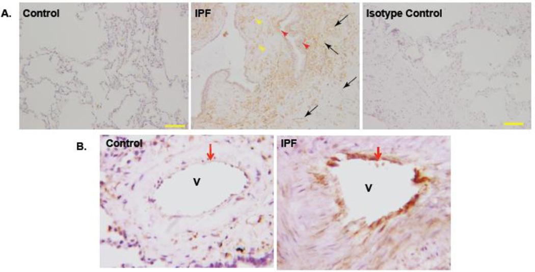
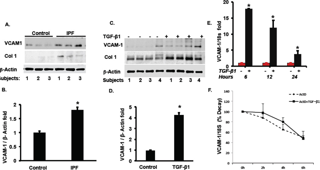
Idiopathic pulmonary fibrosis (IPF) is a chronic lethal interstitial lung disease of unknown etiology. We previously reported that high plasma levels of vascular cell adhesion molecule 1 (VCAM-1) predict mortality in IPF subjects. Here we investigated the cellular origin and potential role of VCAM-1 in regulating primary lung fibroblast behavior. VCAM-1 mRNA was significantly increased in lungs of subjects with IPF compared to lungs from control subjects (p=0.001), and it negatively correlated with two markers of lung function, forced vital capacity (FVC) and pulmonary diffusion capacity for carbon monoxide (DLCO). VCAM-1 protein levels were highly expressed in IPF subjects where it was detected in fibrotic foci and blood vessels of IPF lung. Treatment of human lung fibroblasts with TGF-β1 significantly increased steady-state VCAM1 mRNA and protein levels without affecting VCAM1 mRNA stability. Further, cellular depletion of VCAM-1 inhibited fibroblast cell proliferation and reduced G2/M and S phases of the cell cycle suggestive of cell cycle arrest. These effects on cell cycle progression triggered by VCAM1 depletion were associated with reductions in levels of phosphorylated extracellular regulated kinase 1/2 and cyclin D1. Thus, these observations suggest that VCAM-1 is a TGF-β1 responsive mediator that partakes in fibroblast proliferation in subjects with IPF.

Keywords: IPF; Lung fibroblasts; TGF-β1; VCAM-1.


Conflict of interest statement

Conflicts of interest: None

Figures

Figure 1
Figure 1. VCAM-1 mRNA levels are increased in IPF lungs and negatively correlate with pulmonary function
A. VCAM1 expression levels were extracted from publically available data by microarray profiling of lung tissues from IPF (IPF: n =134) and control subjects (control: n=109). VCAM-1 is up-regulated in IPF subjects compared to control (*P= 0.0001) [22, 23]. B, C: Associations between continuous variables were established by a Spearman correlation analysis and the regression lines have been fitted in two-way scatterplots by Stata software. For FVC association; rho= − 0.2186, P= 0.0123 and for DLco; rho= − 0.2074, P = 0.02. D: VCAM-1 mRNA levels in lungs of control subjects (n=11) and IPF subjects (n=11) were measured using qRT-PCR analysis. Total cellular RNA was extracted with Triazol. The mitochondrial ribosomal protein S18A (MRPS18A) was used as an internal standard. Data represents fold change in VCAM-1/S18A ratios with a ratio for control cells taken as one fold. Data are expressed as means ± SE using an unpaired students t-test *P<0.003 for controls vs IPF groups.
Figure 2
Figure 2. Plasma VCAM-1 levels are increased in IPF subjects
A. Plasma VCAM-1 (ng/ml) was assayed in control subjects (n=50) and IPF (n=48) subjects using an ELISA assay. Approximately equal sample sizes were used in each group and the comparison between each group was computed using an unpaired student t-test. (P = 0.0003, mean = 737.2979 and SD = 186.8587 for IPF and mean = 596.735 and SD = 178.8378 for controls). B, C. Whole lung tissue lysates were obtained from IPF lungs and control lungs as described in Methods. Protein levels of VCAM-1 and collagen 1 (Col1, positive control) were determined in lungs by immunoblotting. Shown is a representative of immunoblot for VCAM1, Col1, and β-actin as a loading control. The bands were quantified by ImageJ and the ratios are presented in panel C showing fold change in IPF lungs compared to control lungs. D. Control mice (C) or bleomycin (BL) treated mice (n=3 mice/group) were euthanized and analyzed for VCAM1 protein in whole lung by immunoblotting (inset) and bands quantitated densitometrically (below).
Figure 3
Figure 3. Expression of VCAM-1 protein in IPF lung
A. Paraffin embedded IPF and control lungs were incubated overnight in primary antibody at 4 ° C. An appropriate secondary antibody was used to identify the target protein on the lung tissue slides. The IPF lung shows staining in endothelial cells (black arrows), airway epithelial cells (red arrowheads) and cells in the interstitium (yellow arrowheads). No staining was detected with the non-immune isotype control (magnification × 100, yellow inset bar=100 microns). B. Brown staining for VCAM-1 protein in IPF lung vessels (V) (arrow) was detected and lack of staining was observed in control lung vessels.
Figure 4
Figure 4. Expression of VCAM-1 protein in primary lung fibroblasts
A–B. Control and IPF human lung fibroblasts were cultured and harvested at 70–80% confluence. VCAM-1 protein was examined by immunoblotting. Collagen 1 served as positive control for IPF lung fibroblasts and β-actin was used as a loading control of individual samples. The immunoblot shown is a representative of three independent experiments. In (B) individual bands were analyzed densitometrically and corrected for loading and densitometric ratios are shown as a bar graph. *P< 0.05 for control vs IPF groups. C–D. Lung fibroblasts were cultured and stimulated with TGF-β1 (5 ng/ml) for 24 hrs. The bands on immunoblots were quantified by ImageJ and densitometric ratios shown in Figure D. Statistical analysis was performed by using an unpaired student t-test. *P= 0.0018 for control vs TGF-β1 groups. Data shown are representatives of 3–5 independent experiments. E. Lung fibroblasts were cultured and stimulated with or without TGF -β1 (E) for 24 hrs. F. Lung fibroblasts were cultured in the presence of actinomycin D (ActD) with or without the inclusion of TGF-β1 (5 ng/ml) in the culture medium for various time points as shown. In both (E) and (F) total cellular RNA was extracted and real time PCR was performed to assay VCAM-1 mRNA as shown. The data represents three separate experiments except panel (F), n=2.
Figure 5
Figure 5. VCAM-1 cellular depletion decreases fibroblast proliferation by impairing cell cycle progression
A. Human lung fibroblasts were transfected with control shRNA or VCAM-1 shRNA and proliferation of cells was assessed using BrdU labeling as described in Methods. Significantly reduced BrdU incorporation was observed in VCAM-1 shRNA transfected cells (*P=< 0.05 in shRNA control vs VCAM-1 shRNA). B. VCAM-1 cellular depletion was conducted as in (A). VCAM-1 silencing on cell cycle progression was then determined in fibroblasts after depleting the adhesion molecule. Significantly reduced S phase and G2/M phase were observed in VCAM-1 shRNA transfected cells (P=< 0.05) versus control shRNA. C–D. Human lung fibroblasts were cultured, transfected and harvested at 70–80% confluence as described above using shRNA. Cell lysates were harvested and processed for immunoblot analysis of indicated cell signaling proteins and key proteins involved in cell cycle regulation. The data are representative of n=2 separate experiments.

References

    1. ATS/ERS International Multidisciplinary Consensus. Classification of the idiopathic interstitial pneumonias. Am J Respir Crit Care Med. 2002;165:277–304. - PubMed
    1. Raghu G CH, Egan JJ, Martinez FJ, Behr J, Brown KK, Colby TV, Cordier JF, Flaherty KR, Lasky JA, Lynch DA, Ryu JH, Swigris JJ, Wells AU, Ancochea J, Bouros D, Carvalho C, Costabel U, Ebina M, Hansell DM, Johkoh T, Kim DS, King TE, Jr, Kondoh Y, Myers J, Müller NL, Nicholson AG, Richeldi L, Selman M, Dudden RF, Griss BS, Protzko SL, Schünemann HJ. An official ATS/ERS/JRS/ALAT statement: idiopathic pulmonary fibrosis: evidence-based guidelines for diagnosis and management. Am J Respir Crit Care Med. 2011;183:788–824. - PMC - PubMed
    1. Lynch J, III, Saggar R, Weigt S, Zisman D, White E. Usual Interstitial Pneumonia. Semin Respir Crit Care Med. 2006;27:634–651. - PubMed
    1. Flaherty K, Travis W, Colby T, Toews G, Kazerooni E, Gross B, Jain A, Strawderman R, III, Flint A, Lynch J, III, Martinez F. Histopathologic variability in usual and nonspecific interstitial pneumonias. Am J Respir Crit Care Med. 2001;164:1722–1727. - PubMed
    1. Selman M, King TE, Pardo A. Idiopathic pulmonary fibrosis: prevailing and evolving hypotheses about its pathogenesis and implications for therapy. Annals of internal medicine. 2001;134:136–151. - PubMed

Publication types

Substances