. 2015 Nov:88:1-13.
doi: 10.1016/j.yjmcc.2015.09.004. Epub 2015 Sep 16.

Cardiomyocyte-specific overexpression of the ubiquitin ligase Wwp1 contributes to reduction in Connexin 43 and arrhythmogenesis

Affiliations

Cardiomyocyte-specific overexpression of the ubiquitin ligase Wwp1 contributes to reduction in Connexin 43 and arrhythmogenesis

Wassim A Basheer et al. J Mol Cell Cardiol. 2015 Nov.

Abstract

Gap junctions (GJ) are intercellular channels composed of connexin subunits that play a critical role in a diverse number of cellular processes in all tissue types. In the heart, GJs mediate electrical coupling between cardiomyocytes and display mislocalization and/or downregulation in cardiac disease (a process known as GJ remodeling), producing an arrhythmogenic substrate. The main constituent of GJs in the ventricular myocardium is Connexin 43 (Cx43), an integral membrane protein that is rapidly turned over and shows decreased expression or function with age. We hypothesized that Wwp1, an ubiquitin ligase whose expression in known to increase in aging-related pathologies, may regulate Cx43 in vivo by targeting it for ubiquitylation and degradation and yield tissue-specific Cx43 loss of function phenotypes. When Wwp1 was globally overexpressed in mice under the control of a β-actin promoter, the highest induction of Wwp1 expression was observed in the heart which was associated with a 90% reduction in cardiac Cx43 protein levels, left ventricular hypertrophy (LVH), and the development of lethal ventricular arrhythmias around 8weeks of age. This phenotype was completely penetrant in two independent founder lines. Cardiomyocyte-specific overexpression of Wwp1 confirmed that this phenotype was cell autonomous and delineated Cx43-dependent and -independent roles for Wwp1 in arrhythmogenesis and LVH, respectively. Using a cell-based system, it was determined that Wwp1 co-immunoprecipitates with and ubiquitylates Cx43, causing a decrease in the steady state levels of Cx43 protein. These findings offer new mechanistic insights into the regulation of Cx43 which may be exploitable in various gap junctionopathies.

Keywords: Arrhythmogenesis; Connexin; Gap junction; Ubiquitin; Wwp1.


Figures

Fig. 1
Fig. 1. Beta-actin driven overexpression of Wwp1 in mice causes sudden death around 8 weeks of age and high levels of expression in the heart
(A) A schematic of the pTraffic construct is depicted. In the inactive state, transgenic mice ubiquitously express DsRed2 resulting in red fluorescence as seen in the representative tail. No fluorescence can be visualized in the green channel. (B) Upon crossing to beta-actin cre transgenic mice, the offspring that inherit both the cre transgene as well as the pTraffic-Wwp1 transgene (tg/+; cre/+) ubiquitously overexpress Wwp1 and GFP as pictured in the tail biopsy and no longer express DsRed2. (C) A Kaplan-Meier plot summarizing the percent survival for 30 Wwp1 overexpressers (tg/+; cre/+) from two independent founder lines (14667 and 14730) along with 30 wild type littermates (+/+; cre/+) from each line observed over a 180 day period is shown. (D) Wwp1 expression was evaluated in a number of tissues derived from four tg/+; cre/+ animals from each line in comparison to four +/+; cre/+ littermates at 8 weeks of age and normalized to Gapdh expression using quantitative real time PCR. SA=splice acceptor, pA=poly-adenylation signal, IRES=internal ribosome entry sequence, BML= bone marrow leukocytes, MSC= mesenchymal stem cells, *p value <0.001.
Fig. 2
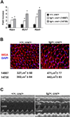
Fig. 2. Mice that globally overexpress Wwp1 develop LVH
(A) Expression of fetal growth genes associated with cardiomyocyte hypertrophy (Nppa, Myh7, and Nppb) was evaluated in the hearts of 8-week old animals using quantitative real time PCR. Wwp1 overexpressers from both lines (14730 and 14667, n=6) exhibited a significant fold increase in the expression levels of Nppa, Myh7 and Nppb when compared to their WT +/+; cre/+ littermates. Mean and standard deviations are shown, *p < 0.01. (B) Wheat germ agglutinin (WGA) staining (in red) of cross sections of the left ventricular myocardium derived from 8 week old 14667 (+/+; cre/+ n=4 and tg/+; cre/+ n= 8) and 14730 mice (+/+; cre/+ n=8 and tg/+; cre/+ n= 7) revealed a significant increase in the cardiomyocyte fiber cross sectional surface area in the left ventricle following Wwp1 overexpression in both lines (*p < 0.001). The calculated mean and standard deviation for the cross sectional area for each line and genotype is shown under their respective images. Scale bars = 10µm. (C) Representative M-mode echocardiographic traces from 8 week old animals (n=4 of each genotype) reveal thickening of both the interventricular septum (IVS) and the left ventricular posterior wall (LVPW) with a concomitant decrease in the left ventricular internal dimension (LVID). Episodic irregular heartbeats were occasionally noted in the Wwp1 overexpressers but never in WT littermates.
Fig. 3
Fig. 3. Wwp1 overexpressers are highly susceptible to ventricular arrhythmias
(A) Langendorff-perfused hearts from 8 week old Wwp1 overexpressors (tg/+; cre/+, n=9) and from their wild type littermates (+/+; cre/+, n=7) were subjected to an overdrive pacing protocol while electrocardiograms were recorded. Shown are typical electrocardiogram traces from these hearts before and after 1.5 sec rapid pacing. (B) In two of the nine hearts derived from Wwp1 overexpressers, spontaneous sustained tachycardia was observed, which was assumed to be lethal in vivo. (C) A summary of the overdrive pacing findings is presented.
Fig. 4
Fig. 4. Hearts derived from animals overexpressing Wwp1 display Cx43 GJ remodeling
(A) Cryostat heart sections derived from 8 week old tg/+; cre/+ animals (n=4) as well as their +/+; cre/+ littermates (n=4) were immunostained for phalloidin (red), N-cadherin (purple) and Cx43 (green). Animals overexpressing Wwp1 showed generalized mislocalization and greatly reduced amounts of Cx43 while expression of N-cadherin was unaffected. Scale bars = 10µm. (B) Frozen sections derived from hearts of 8 week old tg/+; tg/+; cre/+ animals (n=4) or their +/+; cre/+ littermates (n=4) were immunostained for phalloidin (red), desmoplakin (purple) and Cx43 (green). Animals overexpressing Wwp1 showed generalized mislocalization and greatly reduced amounts of Cx43 while expression of desmoplakin was unaffected. Scale bars = 10µm. (C) Representative western blot of total heart protein from 8 week old tg/+; cre/+ and +/+; cre/+ mice illustrating a dramatic decrease in Cx43 protein in both the 14667 and 14730 lines. Normalization and quantification of cardiac Cx43 levels in 8 week old mice revealed a statistically significant 88% reduction in Wwp1 overexpressers from the 14667 line and an 87% decrease in the 14730 line relative to their +/+; cre/+ age matched littermate controls (n=5 for each genotype in each line, *p < 10−6, error bars represent standard deviations). (D) No significant change in Cx43 mRNA levels in the Wwp1 overexpressers (n=6) relative to their +/+; cre/+ littermates (n=6) was detected by qPCR in both lines. Mean and standard deviation are shown.
Fig. 5
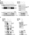
Fig. 5. Wwp1 co-immunoprecipitates with and ubiquitylates Cx43
(A) Cx43 and either MYC-tagged Wwp1 or a MYC-tagged catalytically inactive form of Wwp1 (MYC-C886S) were transfected into 293T cells with either Cx43 or a PY mutant of Cx43 with the proline residue at position 283 mutated to leucine (Cx43-P283L). Both Wwp1 and Wwp1-C886S could be detected in the immunocomplex with WT Cx43; however the interaction between Cx43 and Wwp1 was abrogated when the Cx43-P283L PY mutant was transfected into the cells. Further, co-expression of WT Wwp1 and WT Cx43 resulted in a decrease in the steady state of Cx43 input. (B) To determine whether Wwp1 could ubiquitylate Cx43, 293T cells were transfected with HA-tagged Ub, Cx43, and either WT or MYC-C886S Wwp1 and, 48h later, were stimulated with PMA for 1hr. Five hours after PMA washout, cells were lysed and subjected to Cx43 immunoprecipitation and then the protein extract was immunoblotted with an anti-HA antibody to detect ubiquitylation. While WT Wwp1 mediated robust ubiquitylation of Cx43, the Wwp1-C886S mutant could not. This was also reflected in the decreased Cx43 detected in the input of cells transfected with WT Wwp1 and Cx43.
Fig. 6
Fig. 6. Cardiomyocyte-specific overexpression of Wwp1 yields sudden death and age-dependent LVH
(A) The pTraffic-Wwp1 transgenic mice were mated to mice homozygous for a tamoxifen (Tmx)-inducible cre under the control of the cardiomyocyte-specific alpha -myosin heavy chain promoter (MerCreMer), yielding offspring that carried just the cre construct (+/+; MerCreMer/+) or animals that carried both the pTraffic-Wwp1 and the MerCreMer transgenes (tg/+; MerCreMer/+). Only tg/+; MerCreMer/+ that received two consecutive intraperitoneal injections of Tmx (40 mg/kg/day) beginning at either 26 days of age (n=10) or 54 days of age (n=17) exhibited a sudden cardiac death phenotype around 8 weeks of age (55 and 57 days, respectively). All the vehicle-treated mice (+/+; MerCreMer/+, n=10 for each timepoint or tg/+; MerCreMer/+, n=10 and n=15 for the earlier and later timepoint, respectively) as well as the Tmx-treated +/+; MerCreMer/+ mice (n= 10 for each start point) lived a normal life span as summarized in the Kaplan-Meier plots. (B) Serial transthoracic echocardiographs were obtained from tg/+; MerCreMer mice treated with vehicle or Tmx beginning at 26 days of age (n=3 for each, panels on the left) or at 54 days of age (n=3 for each, panels on the right). Representative M-mode traces illustrate the LVH that developed in Tmx-treated animals 18 days following cre induction but not in vehicle-treated littermates while those tg/+; MerCreMer mice that were administered Tmx (n=3) or vehicle (n=3) beginning at 54 days of age show episodic irregular heartbeats without LVH.
Fig. 7
Fig. 7. Cardiomyocyte-specific overexpression of Wwp1 results in a rapid decrease in Cx43 protein levels, EKG abnormalities and high susceptibility to cardiac arrhythmia
(A) Wwp1 was overexpressed in the cardiomyocytes of tg/+; MerCreMer/+ mice beginning at 26 or 54 days of age via Tmx injection. Total protein was harvested from hearts 72 hours later and Cx43 levels were determined by Western blotting and densitometry. This analysis revealed a 38% decrease in normalized total cardiac Cx43 protein levels in animals injected at 26 days of age and a 45% decrease in normalized total cardiac Cx43 protein levels in animals treated with Tmx beginning at 54 days of age as compared to age-matched, vehicle-injected tg/+; MerCreMer/+ controls (n=3 for each genotype, treatment, and age). (B) Twenty-six day old tg/+; MerCreMer/+ mice were either vehicle- or Tmx-injected (n=3 for each treatment). One week later, hearts from these animals were Langendorff-perfused and subjected to an overdrive pacing protocol while electrocardiograms were recorded. Shown are typical traces from these hearts before and after 1.5 sec rapid pacing. Abnormal baseline waveforms were only noted in the hearts isolated from the Tmx-induced Wwp1 overexpressers. While all vehicle treated hearts quickly recovered sinus rhythm following pacing during this protocol, those hearts with cardiomyocyte specific overexpression of Wwp1 derived from the Tmx-treated tg/+; MerCreMer/+ mice displayed ventricular arrhythmias. (C) Tg/+; MerCreMer/+ mice were injected with either vehicle (n=4) or Tmx (n=4) at 54 days of age and serial electrocardiograms were recorded in this cohort of animals at 12, 24, 48 and 72 hours following injections. Cardiomyocyte-specific overexpression of Wwp1 induced by Tmx caused a significant decrease in the amplitude of the QRS wave at 12 and 24 hours post injection followed by further reduction in the QRS amplitude as well as prolongation of the QT interval at 48 and 72 hours following induction. The vehicle-treated tg/+; MerCreMer/+ mice, on the other hand, showed no significant changes in their EKG traces at any of the time points following injection when compared to the baseline measurement. *p < 0.01

References

    1. Jones SA, Lancaster MK, Boyett MR. Ageing-related changes of connexins and conduction within the sinoatrial node. J Physiol. 2004;560:429–437. - PMC - PubMed
    1. Severs NJ, Bruce AF, Dupont E, Rothery S. Remodelling of gap junctions and connexin expression in diseased myocardium. Cardiovascular research. 2008;80:9–19. - PMC - PubMed
    1. Remo BF, Giovannone S, Fishman GI. Connexin43 cardiac gap junction remodeling: lessons from genetically engineered murine models. The Journal of membrane biology. 2012;245:275–281. - PMC - PubMed
    1. Beardslee MA, Laing JG, Beyer EC, Saffitz JE. Rapid turnover of connexin43 in the adult rat heart. Circulation research. 1998;83:629–635. - PubMed
    1. Darrow BJ, Laing JG, Lampe PD, Saffitz JE, Beyer EC. Expression of multiple connexins in cultured neonatal rat ventricular myocytes. Circulation research. 1995;76:381–387. - PubMed

Publication types

MeSH terms