. 2015 Sep 19:16:60.
doi: 10.1186/s12868-015-0198-7.

RNA interference machinery-mediated gene regulation in mouse adult neural stem cells

Affiliations

RNA interference machinery-mediated gene regulation in mouse adult neural stem cells

Filippo M Cernilogar et al. BMC Neurosci. .

Abstract

Background: Neurogenesis in the brain of adult mammals occurs throughout life in two locations: the subventricular zone of the lateral ventricle and the subgranular zone of the dentate gyrus in the hippocampus. RNA interference mechanisms have emerged as critical regulators of neuronal differentiation. However, to date, little is known about its function in adult neurogenesis.

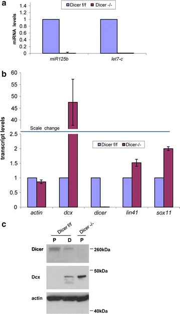
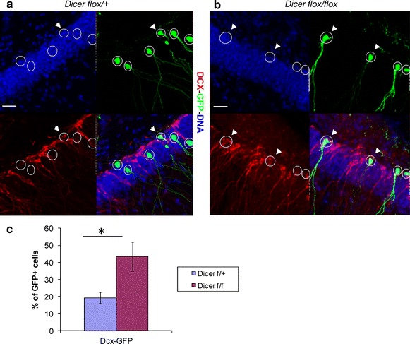
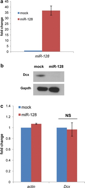
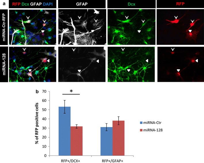
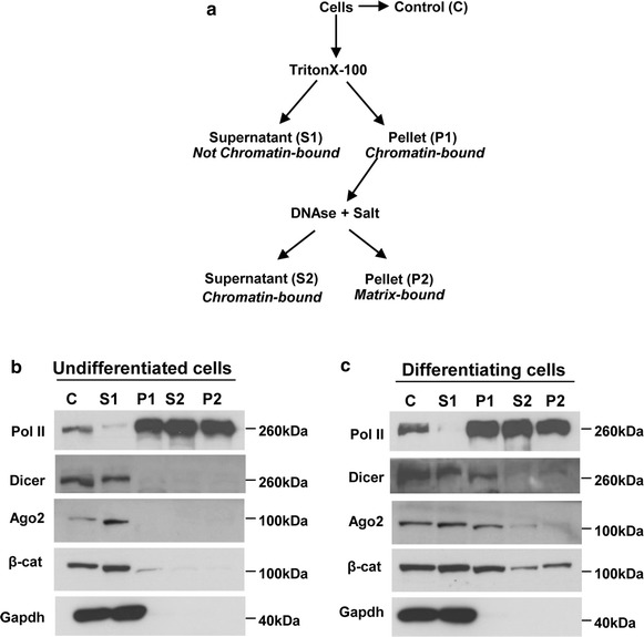
Results: Here we show that the RNA interference machinery regulates Doublecortin levels and is associated with chromatin in differentiating adult neural progenitors. Deletion of Dicer causes abnormal higher levels of Doublecortin. The microRNA pathway plays an important role in Doublecortin regulation. In particular miRNA-128 overexpression can reduce Doublecortin levels in differentiating adult neural progenitors.

Conclusions: We conclude that the RNA interference components play an important role, even through chromatin association, in regulating neuron-specific gene expression programs.


Figures

Fig. 1
Fig. 1
Dcx is up-regulated upon Dicer deletion in adult neural stem cells. ac The samples analyzed were adult neural progenitor cells (NPCs) Dicer flox/flox (f/f) or Dicer −/−. Dicer deletion was obtained by transducing Dicer f/f cells with HTN-Cre protein. a Taqman quantitative RT-PCR of miR125b and let7-c mature miRNA transcripts. Transcript levels were normalized against the housekeeping gene snoRNA55 and calculated respect to Dicer f/f (control) that is set to 1. n = 3, bars represent the mean ± standard error of the mean. b Quantitative RT-PCR of the indicated transcripts. Transcript levels were normalized against the housekeeping gene Srp14 and calculated respect to Dicer f/f (control) that is set to 1. The blue horizontal bar indicates the scale change in the y-axis. n = 3, bars represent the mean ± standard error of the mean. c Western blot showing a representative picture of the Dicer, Dcx and Actin (loading control) protein levels in NPCs (P) or differentiating NPCs (D; 4 days after growth factor withdrawal) of the indicated genotype. Most of the Dicer −/− cells die upon induction of differentiation and therefore are not included in the analysis. Three independent biological samples have been analyzed. Shown are representative pictures
Fig. 2
Fig. 2
Dcx is up-regulated upon Dicer deletion in hippocampal new born neurons. a, b Representative pictures of the immunostaining for Dcx and GFP on hippocampal regions of Dicer cKO mice transduced with Cre-recombinase. a Dicer flox/+ mice (control) 28 dpi. b Dicer flox/flox mice (experiment) 28 dpi. Transduced cells are GFP positive (green) and identified by white circles. Arrowheads indicate Dcx-expressing (red) among the Cre-transduced (green) cells. Single channel and overlay pictures are shown. Scale bar 25 µm. c Quantification of Dcx/GFP double positive cells among the virus-transduced cells. Bars show the average ± the standard error of the mean (n = 4). At least 100 virus-transduced cells per group of four animals were analyzed. Two-tailed t test was applied for statistical analysis. Asterisks indicate statistically relevant differences; p < 0.05
Fig. 3
Fig. 3
Overexpression of miRNA128 reduces Dcx protein levels in N2A cells. ac The samples analyzed were mock-treated Neuro2A cells (control transfection without miRNA-128-RFP expressing plasmid) or Neuro2A cells treated with miRNA-128-RFP expressing plasmid. a Taqman quantitative RT-PCR of the mature miRNA-128 transcripts normalized against the housekeeping small non-coding RNA snoRNA55. Transcript levels are indicated as fold change respect to the control (mock) that is set to 1. n = 3, bars represent the mean ± standard error of the mean. b Western blot showing the Dcx and Gapdh (loading control) protein levels in mock and miRNA-128 over-expressing cells. c Quantitative RT-PCR of the indicated transcripts. Transcript levels are indicated as fold change respect to the control (mock) that is set to 1. Transcripts were normalized against the housekeeping gene Srp14. n = 4, bars represent the mean ± standard error of the mean. Two-tailed t test was applied for statistical analysis. NS not significant; p = 0.4
Fig. 4
Fig. 4
Overexpression of miRNA128 reduces Dcx protein levels in “in vitro” differentiating adult neural stem cells. a, b Adult neural progenitor cells were transfected with miRNA-Ctr-RFP (control) or mirRNA-128-RFP (experiment) expressing plasmids and induced to differentiate by growth factor withdrawal. Analysis was done 6 days later. a Representative pictures of the immunostaining of Dcx, GFAP and RFP on control and experiment cells. DAPI visualizes the DNA. Transfected cells are RFP positive. Arrows indicate Dcx/RFP positive cells; arrowheads indicate GFAP/RFP positive cells. b Quantification of Dcx and GFAP among the transfected cells. Bars show the average ± the standard error of the mean (n = 3). The percentage of marker-positive cells among the total RFP-positive population was calculated on three biological replicates. At least 100 cells per experimental group were analyzed. Two-tailed t test was applied for statistical analysis. Asterisks indicate statistically relevant differences; p < 0.05
Fig. 5
Fig. 5
RNAi components are associated with chromatin in differentiating adult neural stem cells. a Scheme of the procedure followed to fractionate neural progenitor cells. Insoluble fractions (pellets) were dissolved in RIPA buffer (see “Methods”). Chromatin associated proteins should be found in fractions P1 and S2. b, c Equivalent amounts of the resulting protein samples from undifferentiated or differentiating (3 days) adult neural progenitor cells were analyzed by western blot for the presence of the indicated proteins. Gapdh (glyceraldehyde 3-phosphate dehydrogenase) serves as a chromatin unbound marker; Pol II (RNA Polymerase II) is a chromatin bound marker. β-cat β-catenin, Ago2 Argonaute-2. Three independent biological samples have been analyzed. Shown are representative pictures

References

    1. Kokovay E, Shen Q, Temple S. The incredible elastic brain: how neural stem cells expand our minds. Neuron. 2008;60(3):420–429. doi: 10.1016/j.neuron.2008.10.025. - DOI - PubMed
    1. Merkle FT, Alvarez-Buylla A. Neural stem cells in mammalian development. Curr Opin Cell Biol. 2006;18(6):704–709. doi: 10.1016/j.ceb.2006.09.008. - DOI - PubMed
    1. Perrier AL, Studer L. Making and repairing the mammalian brain–in vitro production of dopaminergic neurons. Semin Cell Dev Biol. 2003;14(3):181–189. doi: 10.1016/S1084-9521(03)00010-7. - DOI - PubMed
    1. Zhao C, Deng W, Gage FH. Mechanisms and functional implications of adult neurogenesis. Cell. 2008;132(4):645–660. doi: 10.1016/j.cell.2008.01.033. - DOI - PubMed
    1. Kriegstein A, Alvarez-Buylla A. The glial nature of embryonic and adult neural stem cells. Annu Rev Neurosci. 2009;32:149–184. doi: 10.1146/annurev.neuro.051508.135600. - DOI - PMC - PubMed

Publication types

LinkOut - more resources