. 2015 Oct 1;60(1):47-62.
doi: 10.1016/j.molcel.2015.08.009. Epub 2015 Sep 17.

SPG7 Is an Essential and Conserved Component of the Mitochondrial Permeability Transition Pore

Affiliations

SPG7 Is an Essential and Conserved Component of the Mitochondrial Permeability Transition Pore

Santhanam Shanmughapriya et al. Mol Cell. .

Abstract

Mitochondrial permeability transition is a phenomenon in which the mitochondrial permeability transition pore (PTP) abruptly opens, resulting in mitochondrial membrane potential (ΔΨm) dissipation, loss of ATP production, and cell death. Several genetic candidates have been proposed to form the PTP complex, however, the core component is unknown. We identified a necessary and conserved role for spastic paraplegia 7 (SPG7) in Ca(2+)- and ROS-induced PTP opening using RNAi-based screening. Loss of SPG7 resulted in higher mitochondrial Ca(2+) retention, similar to cyclophilin D (CypD, PPIF) knockdown with sustained ΔΨm during both Ca(2+) and ROS stress. Biochemical analyses revealed that the PTP is a heterooligomeric complex composed of VDAC, SPG7, and CypD. Silencing or disruption of SPG7-CypD binding prevented Ca(2+)- and ROS-induced ΔΨm depolarization and cell death. This study identifies an ubiquitously expressed IMM integral protein, SPG7, as a core component of the PTP at the OMM and IMM contact site.


Figures

Figure 1
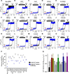
Figure 1. Characterization of the Molecular Identity of Mitochondrial Ca2+-Induced PTP Opening
(A) HEK293T cells stably expressing lentiviral Neg shRNA or shRNA against 128 mitochondrial PTP candidate genes. X-axis numbers represent one PTP candidate knockdown (KD) (see Table S1). Bars represent quantification of the number of [Ca2+]out pulses (10 µM) handled by the mitochondria ([Ca2+]m retention) before collapsing ΔΨm. Mean ± SEM.; n=3–4. See also Figure S1. (B) Representative traces of [Ca2+]out clearance (purple) and ΔΨm (orange). 293T cells stably expressing negative shRNA (control). n=6. See also Figure S1. (C–P) Representative traces of [Ca2+]out clearance (purple) and ΔΨm (orange). 293T cells stably knockdown for PTP candidates sequestered between 14–25 Ca2+ pulses (10 µM) before ΔΨm depolarization. n=3–4. See also Figure S1 and Table S1.
Figure 2
Figure 2. Silencing of SPG7, PPIF, and VDAC1 Prevents Oxidant-Dependent PTP Sensitization
(A–O) Representative traces of [Ca2+]out clearance with or without H2O2 (8 mM) pretreatment at 250 seconds. [Ca2+]out pulses and CCCP were delivered as indicated. 293T cells stably expressing Neg shRNA handled nine Ca2+ pulses (10 µM) without H2O2 pretreatment (black) and three pulses with H2O2 pretreatment (blue). A similar protocol was applied for 14 PTP candidates. n=3–4. (P) Quantification of number of Ca2+ pulses handled in PTP candidate 293T KD cells with (blue) or without H2O2 (black). Mean ± SEM.; n=3–4. (Q) Bar graph illustrates [Ca2+]m retention ratio (R(H2O2+Ca2+)/Ca2+). Mean ± SEM.; n=3–4. See also Figure S2.
Figure 3
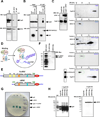
Figure 3. SPG7 Directly Binds CypD
(A) HeLa cell lysate was pretreated with or without CsA (CsA; 5 µM) and immunoprecipitated with SPG7 antibody. IgG was used as a negative control. Inputs (left) and immunoprecipitates (right) were probed with antibodies specific for SPG7 (bottom) and CypD (top). n=5. (B) Western blot analysis of GST-pull down assays performed using purified CypD and GST, SPG7-GST, and AFG3L2-GST. GST protein was used as negative control. Inputs (left) and glutathione sepharose-4B beads pull-down samples (right) were probed with antibodies specific for GST (bottom) and CypD (top). n=4. (C) COS-7 cells expressing CypD-HA, SPG7-HA or CypD-HA and SPG7-HA were lysed, and cell lysates were probed with antibodies specific for HA (left). These cell lysates were separated on a Superdex column, and fractions were immunoblotted with HA antibody (right). COS-7 cells expressing AFG3L2-Flag or AFG3L2-Flag and CypD-Flag were lysed, and cell lysates were fractionated and immunoblotted with Flag antibody. The bottom left panel indicates the coexpression of CypD-Flag and AFG3L2-Flag. n=3. (D) Schematic of yeast two-hybrid assay for SPG7 and CypD interaction. (E) Full-length SPG7 with its functional domains (FL-SPG7;top) and truncated SPG7 lacking N-terminal region (t-SPG7-(Δ270 aa); bottom). (F) Western blot analysis of yeast lysates expressing Myc-tagged Full-length SPG7 (FL-SPG7) and Myc-tagged t-SPG7. n=3. (G) Full-length SPG7 and t-SPG7 were subcloned into GBK vector as bait, and PPIF was cloned into a GAD vector as prey. Upon interaction, yeast expressing GBK-tSPG7/GAD-CypD activated X-α-gal and produced blue colonies (triplicates). GBK-FL-SPG7, GAD-CypD, pGBK-FL-SPG7/pGAD-CypD were negative for X-α-gal. n=3. (H) Western blot analysis of lysates prepared from X-α-gal activation-positive t-SPG7+CypD yeast cells in panel G were immunoblotted for HA (left) and Myc (right). n=3. See also Figure S3.
Figure 4
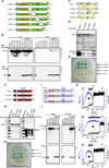
Figure 4. SPG7 C-Terminus Interacts with CypD, and the SPG7 Proteolytic Activity is Dispensable for SPG7-CypD PTP function
(A) Full-length SPG7 with its functional domains (FL-SPG7) and truncated SPG7 (Δ1-Δ5-SPG7). (B) Cell lysates (left) or immunoprecipitated material (right) from COS-7 cells expressing CypD-HA/SPG7-Flag or CypD-HA/Δ1–5-SPG7-Flag and immunoblotted for Flag (Top) and HA (Bottom). n=3. (C) t-SPG7 with its functional domains and truncated t-SPG7 (Δ1-Δ3 t-SPG7) for Y2H assay. (D) Lysates from yeast cells co-expressing CypD-HA/t-SPG7-Myc or CypD-HA/Δ1–3 tSPG7-Myc and immunoblotted for Myc (Top) and HA (Bottom). n=3. (E) As in Figure 3G, yeast were transformed with GBK-t-SPG7/GAD-PPIF and GBK-Δ1–3t–SPG7/GAD-PPIF and subjected to selection. X-α-gal activation was shown as triplicate blue colonies. GBK-Δ3 t-SPG7/GAD-CypD was negative for X-α-gal. n=3. (F) t-SPG7 (top) and t-SPG7 proteolytic domain mutants (Mut1 and Mut2 t-SPG7; bottom). (G) Lysates from yeast cells coexpressing CypD-HA + t-SPG7-Myc, t-SPG7Mut1-Myc, or t-SPG7 Mut2-Myc were immunoblotted for Myc (left) and HA (right). n=3. (H) As in Figure 3G, yeast were transformed with GBK-t-SPG7/GAD-PPIF, GBK-t-SPG7Mut1/GAD-PPIF and GBK-t-SPG7Mut2/GAD-PPIF and subjected to selection. X-α-gal activation was shown as triplicate blue colonies. n=3. (I) Full-length SPG7 (top) and proteolytic domain mutants (Mut1 and Mut2 FL-SPG7; bottom). (J) Cell lysates (left) or immunoprecipitated material (right) from COS-7 cells expressing CypD-HA + FL-SPG7-Flag, SPG7 Mut1-Flag, or SPG7 Mut2-Flag were immunoblotted for Flag (Top) and HA (Bottom). n=4. (K) Similar to Figure 1B, representative traces of [Ca2+]out clearance (black) and ΔΨm (blue). 293T wild-type (top), SPG7 KD (middle) or wild-type cells stably expressing SPG7 Mut2 (bottom). n=3. See also Figure S4.
Figure 5
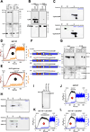
Figure 5. Interaction of CypD, SPG7 and VDAC1 Forms the PTP Complex at OMM and IMM Contact Site
(A) Western blots of cell lysates (left) or immunoprecipitated material (right) from COS-7 cells expressing HA-tagged CypD and/or Flag-tagged SPG7. COS-7 cell lysates were pretreated with CsA (5 µM), immunoprecipitated with HA antibody, and immunoblotted for Flag (top) or HA (bottom). (n = 4). (B) Western blots of cell lysates (left) or immunoprecipitated material (right) from COS-7 cells expressing HA-tagged SPG7, Flag-CypD wild-type, and Flag-CypD-ΔCsA. HA-immunoprecipitated material was immunoblotted for Flag (top) or HA (bottom). (n = 3). (C) COS-7 cells expressing CypD-ΔCsA-Flag (top) or CypD-ΔCsA-Flag and SPG7-Flag (bottom) were lysed, gel filtration column fractions were immunoblotted with Flag antibodies. n=3. (D and E) Similar to Figure 1B, representative traces of [Ca2+]out clearance (orange) and ΔΨm (brown). 293T wild-type (top) or wild-type cells stably expressing CypD-ΔCSA (bottom). n=3. (F) Schematic of full-length SPG7 with its functional domains (FL-SPG7). Mutations in FL-SPG7 are highlighted in red (ΔS1, ΔS2, ΔS3). (G) Western blots of cell lysates (left) or immunoprecipitated material (right) from COS-7 cells expressing SPG7-Flag, VDAC1-HA, and ΔS1-ΔS3-Flag. HA antibody co-immunoprecipitated FL-SPG7-Flag and ΔS2-Flag with VDAC1-HA. n=3. (H) COS-7 cells expressing VDAC1-HA (top) or coexpressing VDAC1-HA and SPG7-HA (bottom) were lysed, and gel filtration column fractions were immunoblotted with HA antibodies. n=3. (I) Western blot analysis of 293T wild-type or wild-type cells stably expressing ΔS1-SPG7-Flag and ΔS2-SPG7-Flag. (J–L) Similar to Figure 1B, representative traces of [Ca2+]out clearance (blue) and ΔΨm (black). 293T wild-type (J), wild-type cells stably expressing ΔS1-SPG7-Flag (K) or ΔS2-SPG7-Flag (K). n=3. See also Figure S5.
Figure 6
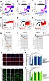
Figure 6. Ca2+ and Oxidant-Induced PTP Opening is a SPG7/PPIF-Dependent Process
(A–C) Representative traces of [Ca2+]out clearance with (blue) or without CSA (pink) (5 µM) pretreatment at 250 seconds. [Ca2+]out pulses and CCCP were delivered as indicated. n=3. (D–F) Mitochondrial membrane integrity in Neg shRNA, SPG7 KD and PPIF KD cells. Permeabilized cells were then challenged with 10 µM Ca2+ bolus pulses after 500 s. Then, MCU blocker Ru360 (1 µM) and CCCP (3 µM) were added as indicated. n=3. (G–I) Isolated mitochondria from Neg shRNA, SPG7 KD and PPIF KD cells were incubated with Ca2+ (250 µM) or t-butyl hydroperoxide (100 µM), and mitochondrial swelling was measured at 540 nm. The mean value traces were plotted with a polynomial fit. n=3–6. (J) Neg shRNA, SPG7 KD and PPIF KD cells were pretreated with DMSO or CsA (5 µM) for 30 min prior to ionomycin (25 µM; 6 hrs) and H2O2 (0.8 mM; 6 hrs) treatment. ΔΨm was measured using TMRE as an indicator. (K) Quantification of mitochondrial TMRE fluorescence. Mean ± SEM; **p<0.01, ***p<0.001, n=3–4. (L) Calcein quenching assay was performed to assess the PTP opening following Ca2+ overload and oxidative stress. Neg shRNA, SPG7 KD and PPIF KD cells were treated as in J. Calcein fluorescence was measured by confocal microscopy. (M) Quantification of mitochondrial calcein fluorescence. Mean ± SEM; **p<0.01, ***p<0.001, n=3–4. See also Figures S6 and S7.
Figure 7
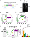
Figure 7. CRISPR/Cas9-Mediated spg7 Knockout Mitochondria Retained High [Ca2+]m and are Resistant to Cell Death
(A) Genotyping strategy for CRISPR/Cas9-Mediated spg7 KO in HEK293T cells. (B) Representative agarose gel image depicts the predicted targeting PCR products. (C and D) Representative traces of [Ca2+]out clearance (magenta) and ΔΨm (green) of wild-type (left) and spg7 knockout 293T cells (right). (E) Western blot depicts the reconstitution of SPG7 Mut2 in spg7 knockout 293T cells. (F) Representative trace of spg7 knockout 293T cells expressing SPG7 proteolytic domain mutant2 (SPG7 Mut2). n=3. (G) Quantification of cell death as PI positive cells. Mean ± SEM; *p<0.05, ***p<0.001, ns, not significant. n=3–4. See also Figures S7.

References

    1. Alavian KN, Beutner G, Lazrove E, Sacchetti S, Park HA, Licznerski P, Li H, Nabili P, Hockensmith K, Graham M, et al. An uncoupling channel within the c-subunit ring of the F1FO ATP synthase is the mitochondrial permeability transition pore. Proc Natl Acad Sci U S A. 2014;111:10580–10585. - PMC - PubMed
    1. Anflous K, Armstrong DD, Craigen WJ. Altered mitochondrial sensitivity for ADP and maintenance of creatine-stimulated respiration in oxidative striated muscles from VDAC1-deficient mice. J Biol Chem. 2001;276:1954–1960. - PubMed
    1. Atorino L, Silvestri L, Koppen M, Cassina L, Ballabio A, Marconi R, Langer T, Casari G. Loss of m-AAA protease in mitochondria causes complex I deficiency and increased sensitivity to oxidative stress in hereditary spastic paraplegia. J Cell Biol. 2003;163:777–787. - PMC - PubMed
    1. Baines CP, Kaiser RA, Purcell NH, Blair NS, Osinska H, Hambleton MA, Brunskill EW, Sayen MR, Gottlieb RA, Dorn GW, et al. Loss of cyclophilin D reveals a critical role for mitochondrial permeability transition in cell death. Nature. 2005;434:658–662. - PubMed
    1. Baines CP, Kaiser RA, Sheiko T, Craigen WJ, Molkentin JD. Voltage-dependent anion channels are dispensable for mitochondrial-dependent cell death. Nat Cell Biol. 2007;9:550–555. - PMC - PubMed

Publication types