Review
. 2016 Jan;238(2):300-10.
doi: 10.1002/path.4641. Epub 2015 Nov 13.

Animal modelling for inherited central vision loss

Affiliations
Review

Animal modelling for inherited central vision loss

Corinne Kostic et al. J Pathol. 2016 Jan.

Abstract

Disease-causing variants of a large number of genes trigger inherited retinal degeneration leading to photoreceptor loss. Because cones are essential for daylight and central vision such as reading, mobility, and face recognition, this review focuses on a variety of animal models for cone diseases. The pertinence of using these models to reveal genotype/phenotype correlations and to evaluate new therapeutic strategies is discussed. Interestingly, several large animal models recapitulate human diseases and can serve as a strong base from which to study the biology of disease and to assess the scale-up of new therapies. Examples of innovative approaches will be presented such as lentiviral-based transgenesis in pigs and adeno-associated virus (AAV)-gene transfer into the monkey eye to investigate the neural circuitry plasticity of the visual system. The models reported herein permit the exploration of common mechanisms that exist between different species and the identification and highlighting of pathways that may be specific to primates, including humans.

Keywords: blindness; cones; fovea; macula; retinal degeneration.


Figures

Figure 1
Figure 1
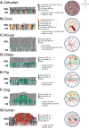
Schematic representation of cone distribution in the central retina of different species. Both sections of the outer nuclear layer and visualization of the eye fundus were schematically represented to highlight the specific cone distribution of the retina of zebrafish, chick, mouse, sheep, pig, dog and human. Photoreceptor types are arbitrarily coloured to represent the different categories of cones (L, M, S, and double cones) and rods. The wavelength of each photopigment is also indicated. (A) In zebrafish, in addition to S‐ and UV‐cones, a double cone is observed with fusion at the level of the IS of two OSs containing either L‐ or M‐opsin. The four classes of cones are then laid out in a regular mosaic pattern. (B) The chick retina contains five types of cones comprising a double cone (in yellow) as well. The different types of cones are homogeneously arranged in the retina. A particular vascular extension, the pecten (red rectangle), is apposed at the inner part of the retina. (C) The mouse retina is composed of only 3% cones, distributed throughout the retina. A dorso‐ventral gradient of cone expression is observed, with S‐opsin mainly expressed in the inferior hemisphere and M‐opsin in the superior hemisphere. However, both opsins are observed in single cells in the overlapping gradient. (D) The sheep retina includes two types of cones, with higher densities of cones in the central streak and in the dorso‐temporal region. S‐cones are enriched in this particular peripheral region. Sheep have a tapetum, a membrane reflecting the light in the superior hemisphere (yellow area) except in the dorso‐nasal periphery. (E) The pig retina features two types of cones, with densities higher in the central streak. (F) The dog retina is also characterized by two types of cones and a central streak, but recently a fovea‐like region was identified with an increased number of cones and a longer OS. Dogs also have a tapetum in the superior hemisphere (yellow area). (G) The human central retina is characterized by a region with exclusively cones named the fovea, containing mainly L‐ and M‐cones. S‐cones are distributed in the perifovea region and in the periphery. ONL: outer nuclear layer; OS: outer segment; black circle: optic nerve head; red lines: vessels; S: superior; I: inferior; N: nasal; T: temporal. Source: refs 32, 72, 94, and 112–127; http://www.cvrl.org/database/text/intros/introdens.htm

References

    1. Hood DC, Finkelstein MA. Sensory processes and perception In Handbook of Perception and Human Performance, Boff KR, Kaufman L, Thomas JP. (eds). John Wiley: Toronto, 1986; 1 : 5‐1–5‐66.
    1. Chew EY, Clemons TE, Sangiovanni JP, et al. Secondary analyses of the effects of lutein/zeaxanthin on age‐related macular degeneration progression: AREDS2 report No. 3. JAMA Ophthalmol 2014; 132 : 142–149. - PMC - PubMed
    1. Petersen‐Jones SM, Komaromy AM. Dog models for blinding inherited retinal dystrophies. Hum Gene Ther Clin Dev 2015; 26: 15–26. - PMC - PubMed
    1. Roosing S, Thiadens AA, Hoyng CB, et al. Causes and consequences of inherited cone disorders. Prog Retin Eye Res 2014; 42: 1–26. - PubMed
    1. Slijkerman RW, Song F, Astuti GD, et al. The pros and cons of vertebrate animal models for functional and therapeutic research on inherited retinal dystrophies. Prog Retin Eye Res 2015; 48 : 137–159. - PubMed

LinkOut - more resources