. 2015 Sep 21:6:8329.
doi: 10.1038/ncomms9329.

Impaired PIEZO1 function in patients with a novel autosomal recessive congenital lymphatic dysplasia

Affiliations

Impaired PIEZO1 function in patients with a novel autosomal recessive congenital lymphatic dysplasia

Viktor Lukacs et al. Nat Commun. .

Abstract

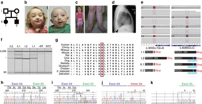
Piezo1 ion channels are mediators of mechanotransduction in several cell types including the vascular endothelium, renal tubular cells and erythrocytes. Gain-of-function mutations in PIEZO1 cause an autosomal dominant haemolytic anaemia in humans called dehydrated hereditary stomatocytosis. However, the phenotypic consequence of PIEZO1 loss of function in humans has not previously been documented. Here we discover a novel role of this channel in the lymphatic system. Through whole-exome sequencing, we identify biallelic mutations in PIEZO1 (a splicing variant leading to early truncation and a non-synonymous missense variant) in a pair of siblings affected with persistent lymphoedema caused by congenital lymphatic dysplasia. Analysis of patients' erythrocytes as well as studies in a heterologous system reveal greatly attenuated PIEZO1 function in affected alleles. Our results delineate a novel clinical category of PIEZO1-associated hereditary lymphoedema.


Figures

Figure 1
Figure 1. PIEZO1 variants in a novel form of hereditary lymphoedema.
(a) Pedigree of the family described in this study. (b) Photographs of II.1 and II.2 at 32 and 7 months of age, respectively. Distinctive facial features include mild infraorbital hypoplasia with subjective hypertelorism, slightly flat facial gestalt with mild periorbital oedema. (c) Photographs of II.1 at 42 months of age. Note abdominal oedema and swelling on the apical surface of the feet. (d) Lateral chest X-ray of II.1 at age 42 months demonstrating pleural effusions. Blunting of the costophrenic angle, a hallmark of pleural effusions, is indicated by an arrow. (e) Alignment of exome sequencing reads (horizontal grey bars) from all individuals to the reference human genome illustrates both the c.3455+1G>A and c.6085G>C mutations. Vertical grey bars next to I.1, I.2, II.1 and II.2 indicate sequencing depth at each base, ∼70 × ; variant bases are highlighted in both the sequencing reads and depth of sequencing bars. The reference sequence and translation are indicated at the bottom of the image. Note that PIEZO1 is encoded by the reverse strand, so variant calls depicted in the sequencing reads are the complement. Predicted protein products for individual family members are depicted below each column of sequencing reads. (f) RT–PCR analysis of patient samples shows defective splicing in those harbouring the c.3455+1G>A mutation. Asterisk indicates shifted product, which is absent in I.2. 5,000 and 50 indicate the internal size standard. (g) Protein alignment of a portion of the C terminus of PIEZO1 illustrating the highly conserved nature of the glycine residue at position 2,029. (h) Sanger sequencing results of RT–PCR products from I.2. (i) Sanger sequencing results of RT–PCR products from I.1. Note the double sequence starting at the exon/intron boundary indicative of retention of intron 24. (j) Deconvolution of dual sequences reveals retention of intron 24 in one allele, with normal splicing from the other allele. (k) Note the software algorithm incorrectly attributes the c.3455+1G>A variant to the exon 25 sequence, so the correct first nucleotide of intron 24 in j is an adenine, while the correct first nucleotide in k is a guanine.
Figure 2
Figure 2. Erythrocytes of patient II.1 display severely decreased PIEZO1 function.
Erythrocytes loaded with Fluo-4 were subjected to mechanical or chemical PIEZO1 stimulation. (a) Schematic of experimental set-up for mechanical stimulation of erythrocytes via negative pipette pressure. (b) Representative response of a control (healthy volunteer) red blood cell to mechanical stimulation, indicated by shaded areas on the trace. Magnitude of negative pressure is indicated above the shaded sections in mm Hg. (c) Average±s.e.m. of responses to negative pipette pressures in four different healthy volunteer individuals are shown as normalized to the response to the calcium ionophore A23187 (4 μM). (d) Average calcium responses of erythrocytes from the four volunteer individuals (depicted in different colours) to the PIEZO1 activator Yoda1 (5 and 15 μM). Cells were stimulated with the compound via whole-chamber perfusion after allowing them to settle and loosely attach to the bottom of the chamber. (e) Red blood cells from I.1, I.2 and II.2 were stimulated mechanically with negative pipette pressures where indicated by shaded grey bars. Numbers above grey bars denote pressure applied in mm Hg. Yoda1 and the calcium ionophore A23187 were applied via whole-chamber perfusion at the end of each measurement. (f) Average±s.e.m. of individual responses normalized to the calcium ionophore A23187 (4 μM). While every parental red blood cell responded to a stimulation greater than −10 mm Hg, no responses were observed to pressures as high as −35 mm Hg in the cells of patient II.2. Statistical comparison of I.1 and I.2 (n=5 in both groups) was performed using the Student's t-test and were found to be not significantly different at the 0.05 level, P=0.21. (g) Red blood cells were stimulated with Yoda1 (15 μM) and the calcium ionophore A23187 (4 μM) as described in d. Background-subtracted Fluo-4 fluorescence intensities from individual cells from I.1, I.2 and II.2 are shown. (h) Average magnitude of calcium responses to 15 μM Yoda1 from g is shown for n=42, 29 and 45 cells of I.1, I.2 and II.2, respectively. One-way analysis was used for statistical comparison. II.2 responses were significantly different from I.1 and I.2, with P values of 2.53E−31 and 1.59E−23, respectively (Bonferroni means comparison test). AFU: arbitrary fluorescence unit.
Figure 3
Figure 3. Attenuated maximal current responses in heterologous cells expressing PIEZO1-G2029R.
Wild-type hPIEZO1 and hPIEZO1-G2029R were expressed in HEK-P1KO cells. (a) Fluorescent calcium imaging of cells loaded with Fura-2 expressing PIEZO1 or PIEZO1-G2029R constructs, both containing EGFP driven by an Internal Ribosomal Entry Site for transfection control. Average fluorescence ratios (340 nm/380 nm) of GFP-positive cells are plotted, normalized to baseline ratio values. (b) Representative whole-cell patch-clamp recordings performed using a blunt-ended mechanical probe, which was moved at 1-μm increments to elicit mechanically induced PIEZO1 and PIEZO1-G2029R current responses. A schematic of the measurement configuration is depicted in the inset. Dashed lines indicate zero current levels in the individual traces. A recording of the displacement steps of the mechanical probe is shown above each trace, with the first step that induced visible contact between the probe and the cell depicted in blue colour. Horizontal scale bars, 25 ms, vertical scale bars, 1 nA. (c) Current–voltage relationships recorded at the 3rd mechano-elicited response show no differences between wild-type and G2029R channels (n=5). Insets show representative recordings for wild-type (black) and G2029R (red) channels. Reversal potential was 3.4±1.08 mV and 3.83±0.45 mV for wild-type and G2029R channels, respectively. Horizontal scale bars, 10 ms, vertical scale bars, 0.1 nA (d) Analysis of mechanically induced current traces shows strongly decreased maximal current densities in the PIEZO1-G2029R channel. Statistics are shown for the mechano-stimulation step 8 μm past where the probe made visible contact (left). Analysis of inactivation and activation kinetics (centre and right, respectively) of the 3rd mechanically induced response (that is, 2 μm past the mechanical threshold) revealed no differences. n=9 and 12 for wild-type and G2029R channels, respectively. Whiskers in the boxplot depict 1.5 interquartile range, star marks indicate range. Statistical analysis was performed using the Mann–Whitney U-test. At the 0.05 level, only current densities were significantly different (P=0.0023).
Figure 4
Figure 4. PIEZO1-G2029R shows decreased surface expression.
HEK-P1KO cells were transfected with either wild-type P1-eMYC or P1-G2029R-eMYC channels. Both constructs contained and extracellular myc tag and an IRES-EGFP for transfection control. (a) Live, unpermeabilized immunofluorescent labelling using an anti-myc antibody show clear surface labelling of 81.5% of the GFP-positive cells in cells expressing P1-eMYC and 17.2% in P1-G2029R-eMYC. (b) Fixed, permeabilized cells were subjected to immunofluorescent labelling with anti-myc and show similar extent of labelling in both wild-type and G2029R groups. Scale bars, 10 μm.

References

    1. Planas-Paz L. et al. Mechanoinduction of lymph vessel expansion. EMBO J. 31, 788–804 (2012). - PMC - PubMed
    1. Sabine A. et al. Mechanotransduction, PROX1, and FOXC2 cooperate to control connexin37 and calcineurin during lymphatic-valve formation. Dev. Cell 22, 430–445 (2012). - PubMed
    1. Coste B. et al. Piezo1 and Piezo2 are essential components of distinct mechanically activated cation channels. Science 330, 55–60 (2010). - PMC - PubMed
    1. Coste B. et al. Piezo proteins are pore-forming subunits of mechanically activated channels. Nature 483, 176–181 (2012). - PMC - PubMed
    1. Ranade S. S. et al. Piezo2 is the major transducer of mechanical forces for touch sensation in mice. Nature 516, 121–125 (2014). - PMC - PubMed

Publication types

MeSH terms

Supplementary concepts