Review
. 2015;88(1055):20150494.
doi: 10.1259/bjr.20150494. Epub 2015 Sep 21.

Total pelvic floor ultrasound for pelvic floor defaecatory dysfunction: a pictorial review

Affiliations
Review

Total pelvic floor ultrasound for pelvic floor defaecatory dysfunction: a pictorial review

Alison J Hainsworth et al. Br J Radiol. 2015.

Abstract

Total pelvic floor ultrasound is used for the dynamic assessment of pelvic floor dysfunction and allows multicompartmental anatomical and functional assessment. Pelvic floor dysfunction includes defaecatory, urinary and sexual dysfunction, pelvic organ prolapse and pain. It is common, increasingly recognized and associated with increasing age and multiparity. Other options for assessment include defaecation proctography and defaecation MRI. Total pelvic floor ultrasound is a cheap, safe, imaging tool, which may be performed as a first-line investigation in outpatients. It allows dynamic assessment of the entire pelvic floor, essential for treatment planning for females who often have multiple diagnoses where treatment should address all aspects of dysfunction to yield optimal results. Transvaginal scanning using a rotating single crystal probe provides sagittal views of bladder neck support anteriorly. Posterior transvaginal ultrasound may reveal rectocoele, enterocoele or intussusception whilst bearing down. The vaginal probe is also used to acquire a 360° cross-sectional image to allow anatomical visualization of the pelvic floor and provides information regarding levator plate integrity and pelvic organ alignment. Dynamic transperineal ultrasound using a conventional curved array probe provides a global view of the anterior, middle and posterior compartments and may show cystocoele, enterocoele, sigmoidocoele or rectocoele. This pictorial review provides an atlas of normal and pathological images required for global pelvic floor assessment in females presenting with defaecatory dysfunction. Total pelvic floor ultrasound may be used with complementary endoanal ultrasound to assess the sphincter complex, but this is beyond the scope of this review.


Figures

Figure 1.
Figure 1.
The normal anatomy during anterior transvaginal scanning. The probe sits in the vagina facing anteriorly to reveal the bladder (B) (dark and large region indicating that it is full of urine), the high-reflective bladder neck (BN) which appears like a white birds' beak, rhabdosphincter (a dark oval area outlined by white dotted line), urethra (U) and the pubic symphysis (PS) in the midsagittal plane.
Figure 2.
Figure 2.
Poor bladder neck support (54-year-old female with stress urinary incontinence) (the ruler along the bottom of each image shows centimetre intervals). The bladder neck (BN) is 3 cm above the pubic symphysis (PS) at rest. During coughing, the BN descends to less than a centimetre above the PS indicating that BN descent is >2 cm. The rhabdosphincter is outlined with a white dotted line, the urethra is seen as the dark thin line marked (U) and the full bladder is marked with a (B).
Figure 3.
Figure 3.
The presence of a transvaginal tape (TVT) (white arrow) in a 45-year-old female (parity 2) presenting with symptoms of an overactive bladder having had a TVT 2 years previously. The synthetic tape is white compared with the surrounding soft tissues. It is lying in the correct position, at the mid-urethra (U). The bladder (B) and pubic symphysis (PS) are marked. BN, bladder neck.
Figure 4.
Figure 4.
Normal posterior transvaginal appearances: the rectum (R), anal canal (A) and anorectal junction (ARJ) are visualized. The puborectalis (PR) appears a white bundle of fibres lying directly behind the rectum, and the perineal body (PB) is a white solid structure, which lies between the probe and the anal canal at the caudal aspect of the image. The internal anal sphincter (IAS) and longitudinal fibres are also marked.
Figure 5.
Figure 5.
Posterior vaginal scans showing intussusception in a 60-year-old female with previous obstetric trauma presenting with obstructive defaecation. There is infolding of the rectal wall (intussusception outlined) during coughing with associated reverberation echoes. The perineal body is marked (PB). Sometimes, less reverberation echoes are seen and the actual infolding is more obvious. If there is poor propulsion or a weak cough, it will not be possible to demonstrate intussusception even if present during defaecation.
Figure 6.
Figure 6.
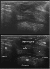
Posterior vaginal scanning shows a rectocoele in a 73-year-old multiparous female presenting with incomplete evacuation requiring vaginal digitation to aid emptying. The arrow shows the bulge of the rectal wall impinging upon the perineal body (PB). The presence of the vaginal probe splints the rectocoele such that it will not protrude any further anteriorly, and the rectocoele appears to be flattened. The puborectalis (PR), rectum (R), anorectal junction (ARJ) and anal canal (A) are also marked. The rectocoele is often only seen during bearing down or coughing, and simultaneous intussusception or infolding of the rectal wall may also be seen.
Figure 7.
Figure 7.
Posterior vaginal scanning demonstrating an enterocoele in a 59-year-old female with post-defaecatory soiling. During bearing down, the enterocoele enters the rectovaginal space and appears as a hyperechoic area between the probe and the rectum (R) such that the rectum is displaced posteriorly. It is not possible to delineate between a sigmoidocoele and enterocoele during ultrasound scanning. The anal canal (A), anorectal junction (ARJ), puborectalis (PR) and perineal body (PB) are also marked.
Figure 8.
Figure 8.
Normal superior endovaginal scan showing the base of bladder anteriorly and rectum posteriorly.
Figure 9.
Figure 9.
A normal endovaginal scan at the upper portion of the urethra. The fibres of the levator ani (LA) and puborectalis (PR) are now visible. The obturator internus (arcuate line insertion of the levators) is also marked.
Figure 10.
Figure 10.
Normal appearances of the endovaginal scan at the level of pubic symphysis (PS) (at 12 o'clock). The pubic arch, deep transverse perineii (deep TP) and puboanalis (PA) are visualized. The symmetry of the pubic arch indicates that the scan is aligned and has been performed correctly with the probe held in a neutral position. The symmetrical insertion of the muscle fibres of the pelvic floor into the pubic arch in both Figures 9 and 10 indicate that the levator plate is intact.
Figure 11.
Figure 11.
The most inferior portion of a normal endovaginal ultrasound scan shows the anal canal and bulbospongiosus (BS).
Figure 12.
Figure 12.
It is useful to assess the symmetry of the cross-sectional images when assessing levator muscle integrity and also to look for the insertion of fibres of the levator ani into the pubic rami on either side. The alignment of the rectum, vagina and urethra is assessed by simply drawing tangential line from the pubic symphysis through these structures. This image shows a levator plate injury in a 43-year-old female presenting with straining, constipation and pelvic pain since giving birth (spontaneous vaginal delivery). There is loss of normal levator muscle integrity on the left (outlined) compared with the right where the levator anatomy is preserved. Consequently, there is a shift of the urethra (small circle) and rectum (large circle) to the left. The pubic arch is still symmetrical, which indicates that the asymmetry visualized is due to levator plate injury rather than rotation of the scan.
Figure 13.
Figure 13.
Endovaginal scan shows a tension-free vaginal tape obturator (TVTO) in place. The synthetic mesh is a white hyperechoic area, looping around the back of the urethra to form a hammock.
Figure 14.
Figure 14.
Normal sagittal transperineal scans: pubic symphysis (PS), bladder, vagina (V), uterus (U), anal canal (A), rectum (R), anorectal angle (ARA) and puborectalis (PR).
Figure 15.
Figure 15.
A rectocoele on sagittal transperineal ultrasound scanning during the Valsalva manoeuvre. The rectum is bulging anteriorly into the vagina. The oval denotes the perineal body (PB). The puborectalis (PR), anal canal (A), anorectal junction (ARJ) and rectum (R) are also marked, and a cystocoele is present.
Figure 16.
Figure 16.
An enterocoele on transperineal scanning. An enterocoele descends from above the rectal ampulla to fill the rectovaginal space, whereas a rectocoele originates from the rectal ampulla and bulges across and over the perineal body (PB). The puborectalis (PR), anal canal (A), anorectal junction (ARJ) and rectum (R) are visualized.
Figure 17.
Figure 17.
A Grade 2 cystocoele is seen as the descent of the bladder into the vagina during coughing in a 56-year-old multiparous patient. The bladder neck is outlined (white bird beak shape) and the bladder (B), vagina (V), rectum (R) and puborectalis (PR) are also marked.

References

    1. Martellucci J, Naldini G. Clinical relevance of transperineal ultrasound compared with evacuation proctography for the evaluation of patients with obstructed defaecation. Colorectal Dis 2011; 13: 1167–72. doi: 10.1111/j.1463-1318.2010.02427.x - DOI - PubMed
    1. Perniola G, Shek C, Chong CC, Chew S, Cartmill J, Dietz HP. Defecation proctography and translabial ultrasound in the investigation of defecatory disorders. Ultrasound Obstet Gynecol 2008; 31: 567–71. doi: 10.1002/uog.5337 - DOI - PubMed
    1. Dietz HP, Beer-Gabel M. Ultrasound in the investigation of posterior compartment vaginal prolapse and obstructed defecation. Ultrasound Obstet Gynecol 2012; 40: 14–27. doi: 10.1002/uog.10131 - DOI - PubMed
    1. Dietz HP. Female pelvic floor dysfunction—an imaging perspective. Nat Rev Gastroenterol Hepatol 2012; 9: 113–21. doi: 10.1038/nrgastro.2011.213 - DOI - PubMed
    1. Beer Gabel M, Assoulin Y, Amitai M, Bardan E. A comparison of dynamic transperineal ultrasound (DTP-US) with dynamic evacuation proctography (DEP) in the diagnosis of cul de sac hernia (enterocoele) in patients with evacuatory dysfunction. Int J Colorectal Dis 2008; 23: 513–19. - PubMed