. 2015 Nov:88:73-81.
doi: 10.1016/j.yjmcc.2015.09.005. Epub 2015 Sep 24.

Characterization of the cardiac succinylome and its role in ischemia-reperfusion injury

Affiliations

Characterization of the cardiac succinylome and its role in ischemia-reperfusion injury

Jennifer A Boylston et al. J Mol Cell Cardiol. 2015 Nov.

Abstract

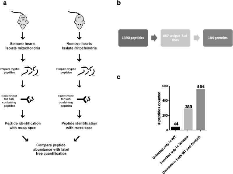
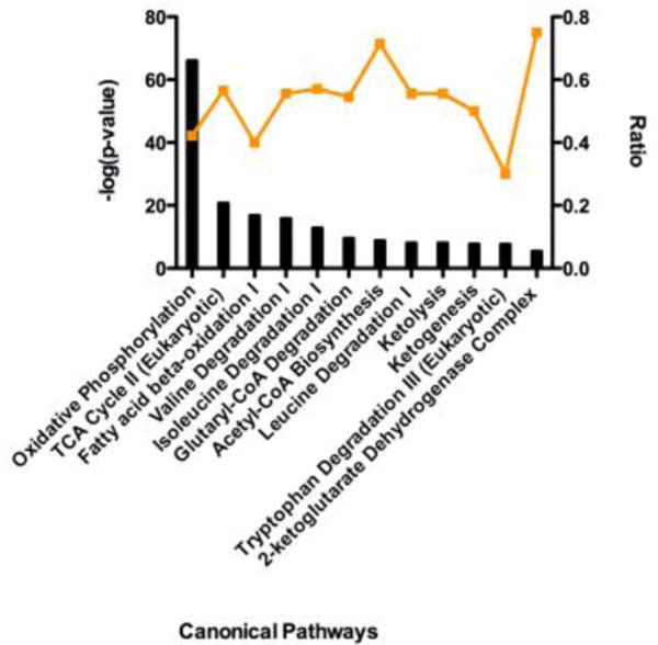
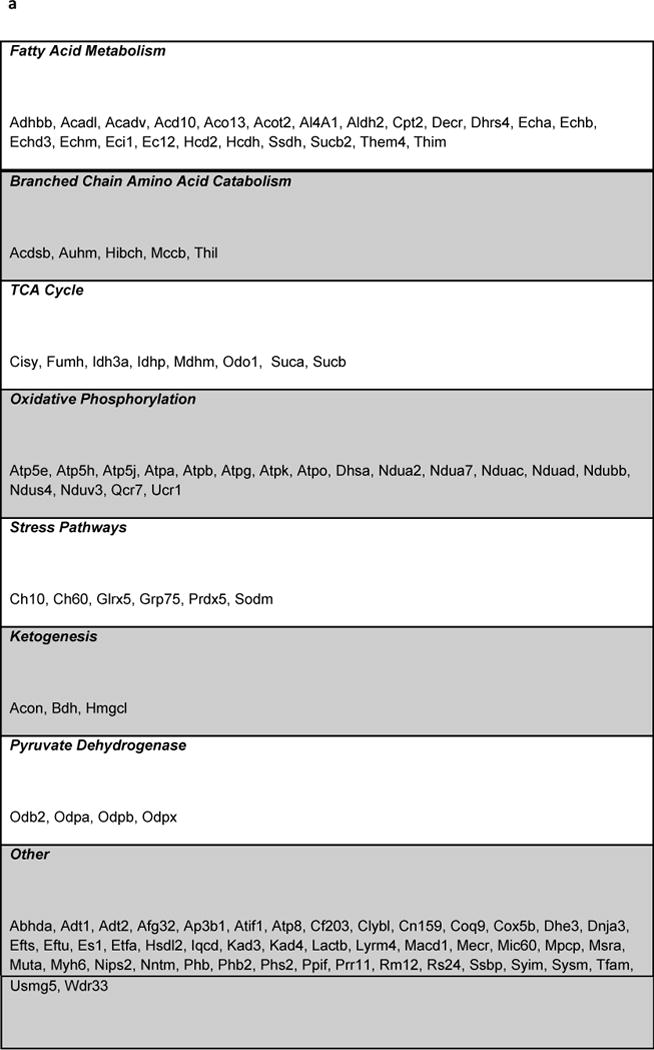
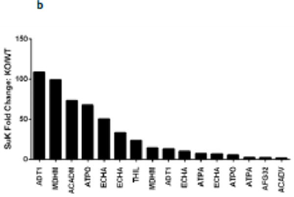
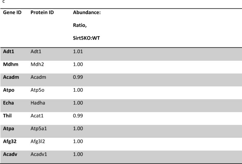
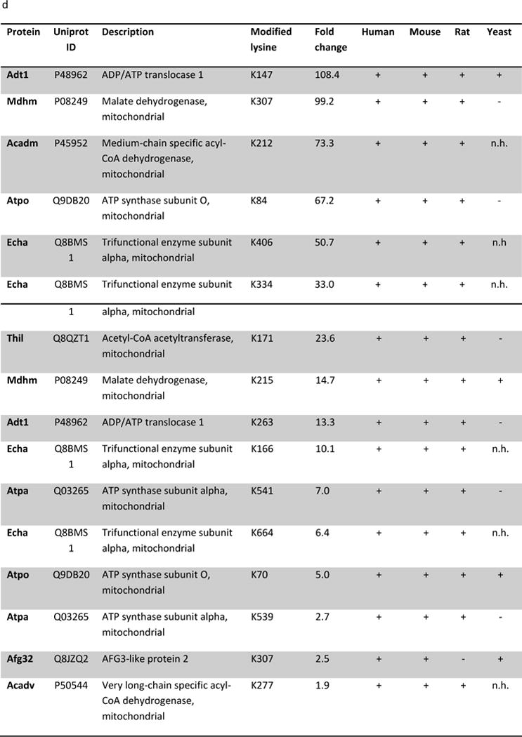
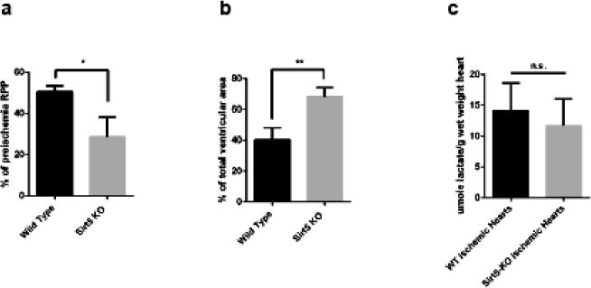
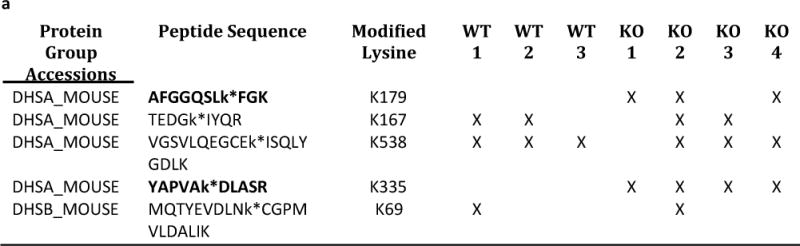
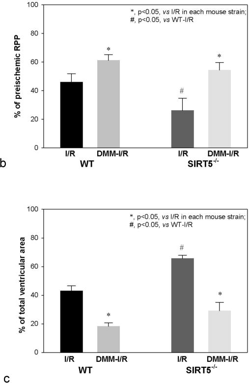
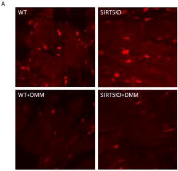
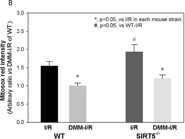
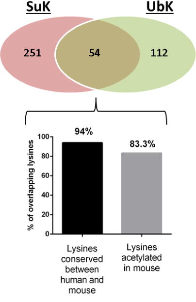
Succinylation refers to modification of lysine residues with succinyl groups donated by succinyl-CoA. Sirtuin5 (Sirt5) is a mitochondrial NAD(+)-dependent deacylase that catalyzes the removal of succinyl groups from proteins. Sirt5 and protein succinylation are conserved across species, suggesting functional importance of the modification. Sirt5 loss impacts liver metabolism but the role of succinylation in the heart has not been explored. We combined affinity enrichment with proteomics and mass spectrometry to analyze total succinylated lysine content of mitochondria isolated from WT and Sirt5(-/-) mouse hearts. We identified 887 succinylated lysine residues in 184 proteins. 44 peptides (5 proteins) occurred uniquely in WT samples, 289 (46 proteins) in Sirt5(-/-) samples, and 554 (133 proteins) were common to both groups. The 46 unique proteins in Sirt5(-/-) heart participate in metabolic processes such as fatty acid β-oxidation (Eci2) and branched chain amino acid catabolism, and include respiratory chain proteins (Ndufa7, 12, 13, Dhsa). We performed label-free analysis of the peptides common to WT and Sirt5(-/-) hearts. 16 peptides from 9 proteins were significantly increased in Sirt5(-/-) by at least 30%. The adenine nucleotide transporter 1 showed the highest increase in succinylation in Sirt5(-/-) (108.4 fold). The data indicate that succinylation is widespread in the heart and enriched in metabolic pathways. We examined whether the loss of Sirt5 would impact ischemia-reperfusion (I/R) injury and we found an increase in infarct size in Sirt5(-/-) hearts compared to WT littermates (68.5(+)/-1.1% Sirt5(-/-) vs 39.6(+)/(-) 6.8% WT) following 20min of ischemia and 90-min reperfusion. We further demonstrate that I/R injury in Sirt5(-/-) heart is restored to WT levels by pretreatment with dimethyl malonate, a competitive inhibitor of succinate dehydrogenase (SDH), implicating alteration in SDH activity as causative of the injury.

Keywords: Cardiac; Ischemia–reperfusion; Sirt5; Succinate; Succinylation.


Figures

Figure 1
Figure 1. Identification of sites of lysine succinylation in cardiac mitochondria
(a) Mouse heart mitochondria were isolated from Sirt5−/− (n=4) and WT (n=3) mice. Each sample was digested with trypsin and succinylated lysine (SuK) containing peptides were isolated by immunoprecipitation with an antibody designed to recognize SuK (PTM Biolabs, PTM-401). Isolated peptides were analyzed with mass spectrometry and label free analysis was used to quantify the relative abundance of peptides common between the sample groups. (b) To be considered for analysis, a peptide must have been detected in at least 2 of the 7 samples. We identified 1390 succinylated (SuK) peptides, which corresponded to 887 unique sites of lysine succinylation. These 887 peptides constitute the cardiac succinylome, and map to 184 proteins. (c) Of the 887 identified SuK peptides, 44 occurred uniquely in WT samples, 289 occurred uniquely in Sirt5 KO samples, and 554 were common to both groups.
Figure 2
Figure 2. Pathways enriched with succinylated proteins
IPA core analysis was used to generate a list of pathways enriched with succinylated proteins. The top 12 scoring canonical pathways are represented. The left y-axis bar represents the [−log (p-value)] of the pathway, calculated by Fisher’s exact test. The right y-axis points (orange line) represent the ratio calculated by dividing the number of succinylated proteins that map to a particular pathway by the total number of proteins in that pathway. The top scoring pathways include oxidative phosphorylation, the TCA cycle, and β-oxidation of fatty acids.
Figure 3
Figure 3. Identification of Sirt5 substrates
(a) IPA core analysis was used to functionally classify proteins mapped from peptides that were detected only Sirt5−/− samples. (b) A label free analysis program (QUOIL) was used to determine the relative abundance of succinylated peptides detected in both WT and Sirt5−/− samples. Represented here are the 16 peptides from 9 proteins increased by > 30% with p < 0.05 in Sirt5−/− hearts. (c) Relative protein quantitation was calculated based on the intensities of TMT report ions using Scaffold 4.0 (Proteome Software, Portland, OR). The average ratio of protein expression (Sirt5−/−: WT) is expressed numerically. The data demonstrate no significant differences in total protein abundance between Sirt5−/− and WT mice. (d) Conservation of the Sirt5 substrate lysines across human, mouse, rat, and S. Cerevisiae (yeast). Protein alignments were performed with the “Align” tool at www.uniprot.org; “+” indicates a conserved lysine, “−“ indicates that the lysine is not conserved, “n.h.” indicates that a homologous protein is not expressed by the organism.
Figure 4
Figure 4. Sirt5−/− mice are susceptible to ischemia-reperfusion injury
(a) Assessment of the rate pressure product (RPP = heart rate*systolic blood pressure) after ischemia-reperfusion injury in hearts of three female WT and three female Sirt5−/− mice. Baseline RPP priiror to ischemia averaged 54,009+/−4572 (HR*cm of water), and LVDP at baseline was 130+/−9 cm of water. (b) Infarct size in WT and Sirt5−/− mice following 20 minutes of global ischemia and 90 minutes of reperfusion. (c) WT and Sirt5−/− mice show no difference in production of lactate after 20 minutes of global ischemia *p<0.05; **p<0.01; n.s., not significant, measured by t-test. Values are means +/− SEM n=3 in each group.
Figure 5
Figure 5. Inhibition of succinate dehydrogenase attenuates injury in WT hearts and restores recovery in Sirt5−/− hearts
(a) M/S identification of 5 peptides that map to protein components of the SDH complex. 2/4 peptides mapped to SDH subunit Dhsa (bold font) are classified as Sirt5 substrates, as these peptides were only detected in Sirt5−/− samples. (b) Assessment of the rate pressure product (RPP = heart rate*systolic blood pressure) when a competitive inhibitor of SDH, dimethyl malonate, is delivered to WT and Sirt5−/− hearts before ischemia-reperfusion injury. Malonate, which is generated in the cell upon addition of dimethyl malonate attenuates injury in the WT heart and restores recovery in Sirt5−/− hearts to WT levels. Baseline RPP prior to ischemia (or addition of DMM) was 45,585+/−1922, and baseline LVDP was 124+/−4 cm of water. Significance was determined by ANOVA followed by a post hoc Tukey.t-test. Values are means +/− SEM. N=11 for WT I/R (no drug), n=6 for WT I/R treated with dimethyl malonoate (DMM-I/R), n=9 for SIRT5KO (no drug), and n=4 for SIRT5KO + DMM-I/R. Male and female mice were used in this study. There were no sex differences. (c) Infarct size in WT and Sirt5−/− mice after hearts were pretreated with 5 mM methyl malonate for 20 minutes before 20 minutes of global ischemia and 90 minutes of reperfusion. Significance was determined by ANOVA followed by a post hoc. Values are means +/− SEM.N values are the same as 5b.
Figure 6
Figure 6. Mito Sox measurement of superoxide in I/R
Panel A shows representative images of section of WT, SIRT5-KO, WT + dimethyl malonate (DMM) and SIRT5-KO + DMM hearts treated with Mito Sox following 20 min of ischemia and 5 min of reperfusion. The bright red spots are due to Mito Sox binding to nucleotides as confirmed by co-localization with DAPI (data not shown). The mean and standard error for each group (n=4) are provided in panel B. * indicates Ssignificantly was determined difference between groups as measured by Kruskal-Wallis ANOVA and a Student-Newman Keuls test.
Figure 7
Figure 7. Overlap of succinylation and ubiquitination in mouse heart
The Venn diagram illustrates the overlap between Sirt5 targeted lysines and ubiquitination. Of the 54 overlapping lysine residues, 51/54 (94%) are conserved between mouse and human. 45/54 (83.3%) are known to be acetylated in mouse.

Comment in

References

    1. Zhao S, Xu W, Jiang W, Yu W, Lin Y, Zhang T, et al. Regulation of cellular metabolism by protein lysine acetylation. Science. 2010;327:1000–4. - PMC - PubMed
    1. Kim SC, Sprung R, Chen Y, Xu Y, Ball H, Pei J, et al. Substrate and functional diversity of lysine acetylation revealed by a proteomics survey. Mol Cell. 2006;23:607–18. - PubMed
    1. Peng C, Lu Z, Xie Z, Cheng Z, Chen Y, Tan M, et al. The first identification of lysine malonylation substrates and its regulatory enzyme. Mol Cell Proteomics. 2011;10:M111 012658. - PMC - PubMed
    1. Zhang Z, Tan M, Xie Z, Dai L, Chen Y, Zhao Y. Identification of lysine succinylation as a new post-translational modification. Nat Chem Biol. 2011;7:58–63. - PMC - PubMed
    1. Tan M, Peng C, Anderson KA, Chhoy P, Xie Z, Dai L, et al. Lysine glutarylation is a protein posttranslational modification regulated by SIRT5. Cell Metab. 2014;19:605–17. - PMC - PubMed

Publication types

MeSH terms