Review
. 2015 Dec:36:13-18.
doi: 10.1016/j.dnarep.2015.09.003. Epub 2015 Sep 10.

Nucleotide excision repair in humans

Affiliations
Review

Nucleotide excision repair in humans

Graciela Spivak. DNA Repair (Amst). 2015 Dec.

Abstract

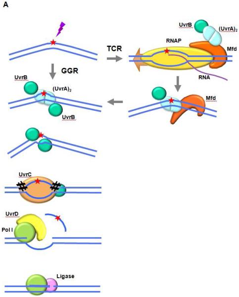
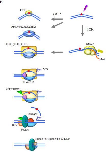
The demonstration of DNA damage excision and repair replication by Setlow, Howard-Flanders, Hanawalt and their colleagues in the early 1960s, constituted the discovery of the ubiquitous pathway of nucleotide excision repair (NER). The serial steps in NER are similar in organisms from unicellular bacteria to complex mammals and plants, and involve recognition of lesions, adducts or structures that disrupt the DNA double helix, removal of a short oligonucleotide containing the offending lesion, synthesis of a repair patch copying the opposite undamaged strand, and ligation, to restore the DNA to its original form. The transcription-coupled repair (TCR) subpathway of NER, discovered nearly two decades later, is dedicated to the removal of lesions from the template DNA strands of actively transcribed genes. In this review I will outline the essential factors and complexes involved in NER in humans, and will comment on additional factors and metabolic processes that affect the efficiency of this important process.

Keywords: DNA repair; Global genome repair; Nucleotide excision repair; Transcription-coupled repair.


Figures

Fig. 1
Fig. 1. Schematics of NER in the prototypic bacterium E. coli, and in humans
A. NER in E. coli The UvrAB complex binds to the DNA and recognizes and verifies the damage to be repaired. UvrA dissociates from the preincision complex leaving one or two molecules of UvrB bound to the DNA. UvrC interacts with UvrB and catalyzes two nicks in the DNA, one on either side of the lesion. The combined action of UvrD (helicase I) and DNA polymerase I removes the oligonucleotide containing the lesion, as well as UvrB and UvrC, from the DNA and results in the synthesis of a patch using the undamaged complementary strand as a template. DNA ligase I seals the patch to the contiguous DNA strand. In TCR, Mfd is activated by binding to a stalled RNAP. It dissociates the RNAP and nascent transcript from the DNA and recruits UvrA complexed with UvrB. Repair then proceeds through the same reaction sequence as GGR. B. NER in humans Higher eukaryotes utilize different mechanisms for detecting DNA alterations in actively transcribed genes and in the genome as a whole. In TCR, RNAPII is arrested at a lesion. TCR factors are recruited; the polymerase is removed or backtracked to allow access to TFIIH and other NER repair enzymes. In GGR, a helix distorting lesion or structure can be directly recognized by XPC complexed with hRAD23B and centrin 2 (CETN2). Certain lesions such as CPD, which do not significantly destabilize DNA duplexes, are first recognized by DDB2 (XPE) in complex with DDB1, creating a kink that is recognized by XPC. The XPC-hRAD23b-CETN2 complex melts the DNA around the lesion and attracts the multiunit complex TFIIH. TCR and GGR converge; XPB and XPD unwind the DNA to create a ~30-nucleotide bubble. Once the pre-incision complex is assembled, XPA, RPA and XPG are recruited and the XPC complex is released. XPA binds the DNA near the 5′ side of the bubble, and RPA binds the ssDNA opposite the lesion, protecting it from degradation and coordinating excision and repair events. XPG and ERCC1-XPF associate with TFIIH. ERCC1-XPF makes the first incision, and repair synthesis proceeds for several nucleotides displacing the damaged strand; XPG incises the 3’ single/double strand junction, and ligase I or ligase IIIα-XRCC1 seal the DNA.

References

    1. Spivak G, Ganesan AK. The complex choreography of transcription-coupled repair. DNA Repair (Amst) 2014;19:64–70. - PubMed
    1. Ganesan A, Spivak G, Hanawalt PC. Transcription-coupled DNA repair in prokaryotes. Prog Mol Biol Transl Sci. 2012;110:25–40. - PubMed
    1. Scharer OD. Nucleotide excision repair in eukaryotes. Cold Spring Harb Perspect Biol. 2013;5(10):a012609. - PMC - PubMed
    1. Marteijn JA, et al. Understanding nucleotide excision repair and its roles in cancer and ageing. Nature Reviews Molecular Cell Biology. 2014;15(7):465–481. - PubMed
    1. Oh DH, Spivak G. Hereditary photodermatoses. Adv Exp Med Biol. 2010;685:95–105. - PubMed

LinkOut - more resources