. 2015 Oct 6;6(30):30263-76.
doi: 10.18632/oncotarget.4749.

Nuclear HDAC6 inhibits invasion by suppressing NF-κB/MMP2 and is inversely correlated with metastasis of non-small cell lung cancer

Affiliations

Nuclear HDAC6 inhibits invasion by suppressing NF-κB/MMP2 and is inversely correlated with metastasis of non-small cell lung cancer

Chih-Jen Yang et al. Oncotarget. .

Abstract

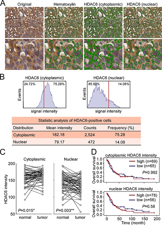
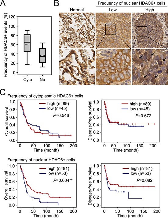
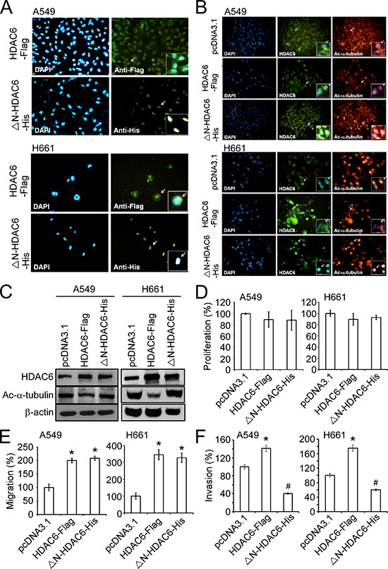
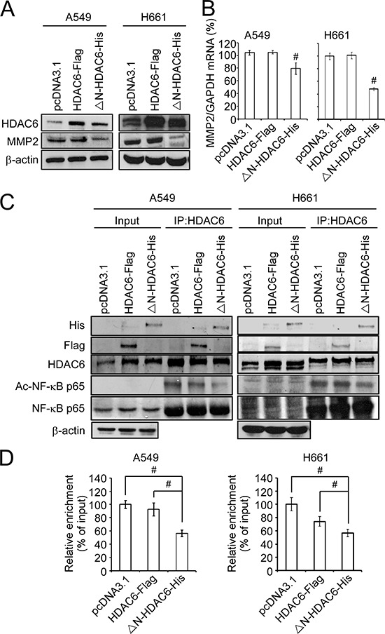
Histone deacetylase 6 (HDAC6) is a unique member of the histone deacetylase family. Although HDAC6 is mainly localized in the cytoplasm, it can regulate the activities of the transcription factors in the nucleus. However, a correlation of intracellular distribution of HDAC6 with tumor progression is lacking. In this study, we found that a low frequency of nuclear HDAC6-positive cells in tumors was associated with distant metastasis and a worse overall survival in 134 patients with non-small cell lung cancer (NSCLC). Ectopic expression of wild-type HDAC6 promoted migration and invasion of A549 and H661 cells. However, the enforced expression of nuclear export signal-deleted HDAC6 inhibited the invasion but not the migration of both cell lines. The inhibitory effect of nuclear HDAC6 on invasion was mediated by the deacetylation of the p65 subunit of nuclear factor-κB, which decreased its DNA-binding activity to the MMP2 promoter, leading to the downregulation of MMP2 expression. Our findings indicated that the loss of nuclear HDAC6 may be a potential biomarker for predicting metastasis in patients with NSCLC.

Keywords: HDAC6; MMP2; NF-κB; lung cancer; metastasis.


Conflict of interest statement

CONFLICTS OF INTEREST

The authors declare no conflict of interest.

Figures

Figure 1
Figure 1. Analysis of HDAC6 cytoplasmic and nuclear localization
A. Representative image showed the identification of hematoxylin-stained nucli and cytoplasmic and nuclear HDAC6 in a section of paraffin-embedded NSCLC specimen using HistoQuest software. B. Quantitative analysis of the intensity and frequency of cytoplasmic and nuclear HDAC6. Red line indicated the 50% cutoff of the HDAC6 intensity. C. The signal intensity of cytoplasmic and nuclear HDAC6 in the paired normal tissues and tumors in 63 NSCLC patients was plotted. *P < 0.05, **P < 0.01. D. Kaplan-Meier log-rank analysis for the overall survival of the 134 NSCLC patients with low and high intensity of cytoplasmic and nuclear HDAC6 in the tumor regions.
Figure 2
Figure 2. The association of nuclear HDAC6 frequency with NSCLC patient survival
A. The frequency (%) of cytoplasmic and nuclear HDAC6 in the tumor parts of the 134 NSCLC patients. Cyto, cytoplasmic HDAC6; Nu, nuclear HDAC6. B. The representative images demonstrated the low and high frequency of nuclear HDAC6+ cells in tumor regions. Images of HDAC6 staining in two normal tissues were shown for comparison. C. Kaplan-Meier log-rank analysis for the overall and disease-free survival of the 134 NSCLC patients with low and high frequency of cytoplasmic (upper panels) and nuclear (lower panels) HDAC6 in the tumor regions. **P < 0.01.
Figure 3
Figure 3. Nuclear expression of HDAC6 inhibited invasion of NSCLC cell lines
A. Immunofluorescence images showed the intracellular localization of HDAC6-Flag and ΔN-HDAC6-His using the antibodies against Flag and His tag (green) in A549 (upper panels) and H661 (lower panels) cell lines. The nucli were stained by DAPI (blue). Arrows indicated the cells shown in the inserts with higher magnification. B. Immunofluorescence images showed the intracellular localization of HDAC6-Flag or ΔN-HDAC6-His (middle panels; green) and acetylated α-tubulin (right panels; red) in A549 (upper panels) and H661 (lower panels) cell lines. The nucli were stained by DAPI (left panels; blue). Arrows indicated the cells shown in the inserts with higher magnification. C. A549 and H661 cells were transfected with pcDNA3.1, HDAC6-Flag or ΔN-HDAC6-His, and the expression of HDAC6, acetylated α-tubulin and β-actin were analyzed by Western blot. D. The cell proliferation of pcDNA3.1-, HDAC6-Flag- or ΔN-HDAC6-His-transfected A549 and H661 cells were analyzed by XTT assay. E. The migration of pcDNA3.1-, HDAC6-Flag- or ΔN-HDAC6-His-transfected A549 and H661 cells were analyzed by wound-healing assay. *P < 0.05, compared with the pcDNA3.1 control. F. The invasion of pcDNA3.1-, HDAC6-Flag- or ΔN-HDAC6-His-transfected A549 (left) and H661 (right) cells were analyzed by transwell invasion assay. *P < 0.05, up-regulation, compared with the pcDNA3.1 control. #P < 0.05, down-regulation, compared with the pcDNA3.1 control.
Figure 4
Figure 4. Nuclear HDAC6 inhibits MMP2 through de-acetylating NF-κB p65 subunit
A. A549 and H661 cells were transfected with pcDNA3.1, HDAC6-Flag or ΔN-HDAC6-His, and the expression of HDAC6, MMP2 and β-actin were analyzed by Western blot. B. The mRNA expression of MMP2 was analyzed by real-time PCR. C. The interaction of HDAC6 and NF-κB was analyzed by immunopreciptation using the antibody against HDAC6 in pcDNA3.1-, HDAC6-Flag- or ΔN-HDAC6-His-transfected A549 (left) and H661 (right) cells. D. Chromatin immunoprecipitation was performed to the pcDNA3.1-, HDAC6-Flag- or ΔN-HDAC6-His-transfected A549 (left) and H661 (right) cells. Real-time PCR was performed using the primers for NF-κB binding sites in MMP2 promoter.

Similar articles

Cited by

References

    1. Yang PH, Zhang L, Zhang YJ, Zhang J, Xu WF. HDAC6: physiological function and its selective inhibitors for cancer treatment. Drug Discov Ther. 2013;7:233–242. - PubMed
    1. Verdel A, Khochbin S. Identification of a new family of higher eukaryotic histone deacetylases. Coordinate expression of differentiation-dependent chromatin modifiers. The Journal of biological chemistry. 1999;274:2440–2445. - PubMed
    1. Grozinger CM, Hassig CA, Schreiber SL. Three proteins define a class of human histone deacetylases related to yeast Hda1p. Proceedings of the National Academy of Sciences of the United States of America. 1999;96:4868–4873. - PMC - PubMed
    1. Bertos NR, Gilquin B, Chan GK, Yen TJ, Khochbin S, Yang XJ. Role of the tetradecapeptide repeat domain of human histone deacetylase 6 in cytoplasmic retention. J Biol Chem. 2004;279:48246–48254. - PubMed
    1. Aldana-Masangkay GI, Sakamoto KM. The role of HDAC6 in cancer. J Biomed Biotechnol. 2011;2011:875824. - PMC - PubMed

Publication types

MeSH terms