Review
. 2015 Sep 1:9:296.
doi: 10.3389/fnins.2015.00296. eCollection 2015.

The role of cerebellar circuitry alterations in the pathophysiology of autism spectrum disorders

Affiliations
Review

The role of cerebellar circuitry alterations in the pathophysiology of autism spectrum disorders

Matthew W Mosconi et al. Front Neurosci. .

Abstract

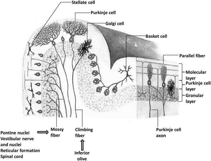
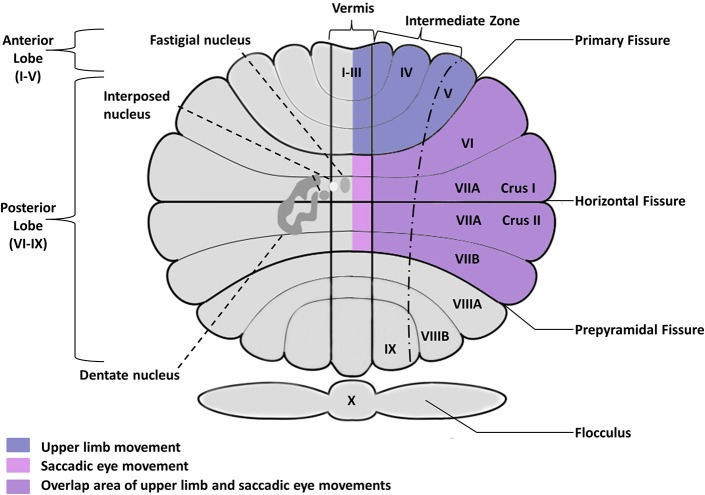
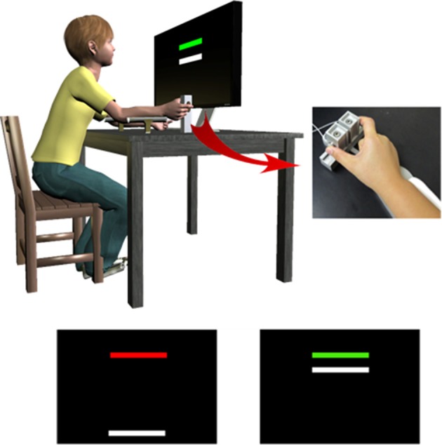
The cerebellum has been repeatedly implicated in gene expression, rodent model and post-mortem studies of autism spectrum disorder (ASD). How cellular and molecular anomalies of the cerebellum relate to clinical manifestations of ASD remains unclear. Separate circuits of the cerebellum control different sensorimotor behaviors, such as maintaining balance, walking, making eye movements, reaching, and grasping. Each of these behaviors has been found to be impaired in ASD, suggesting that multiple distinct circuits of the cerebellum may be involved in the pathogenesis of patients' sensorimotor impairments. We will review evidence that the development of these circuits is disrupted in individuals with ASD and that their study may help elucidate the pathophysiology of sensorimotor deficits and core symptoms of the disorder. Preclinical studies of monogenetic conditions associated with ASD also have identified selective defects of the cerebellum and documented behavioral rescues when the cerebellum is targeted. Based on these findings, we propose that cerebellar circuits may prove to be promising targets for therapeutic development aimed at rescuing sensorimotor and other clinical symptoms of different forms of ASD.

Keywords: autism spectrum disorder; cerebellum; gait; genetics; oculomotor; pathophysiology; precision grip; sensorimotor.


Figures

Figure 1
Figure 1
The microstructural organization of the cerebellar cortex showing the presence of three layers and the relative position of Purkinje, basket, stellate, and Golgi cells and the main inputs (mossy and climbing fibers) of the cerebellum. There are two main afferents to the cerebellar cortex: climbing fibers, which make direct excitatory contact with the Purkinje cells, and mossy fibers, which terminate in the granular layer and make excitatory synaptic contacts primarily with granule cells, but also with Golgi cells. The ascending axons of the granule cells branch in a T-shaped manner to form the parallel fibers, which, in turn, make excitatory synaptic contacts with Purkinje cells and molecular layer interneurons including stellate and basket cells.
Figure 2
Figure 2
Posterior view of the human cerebellum, showing the cerebellar fissure, lobular organization, and deep nuclei embedded within the cerebellar cortex. Deep nuclei are located bilaterally but shown only in the left hemisphere for clarity purposes. Saccadic and smooth pursuit eye movements are controlled by the oculomotor vermis including posterior lobules VI–VII, Crus I–II of the ansiform lobule, and their outputs in caudal fastigial nuclei. Upper limb movements primarily involve anterior lobules I–V as well as more lateral areas of lobules V–VI extending into Crus I–II. Cerebellar circuits involved in controlling balance and gait have been identified in the vermis and intermediate cerebellum (not shown).
Figure 3
Figure 3
To assess precision gripping control during rise, sustained, and relaxation phases, individuals pressed against two opposing load cells while receiving visual feedback from the monitor in front of them. Individuals viewed two horizontal bars: a red/green target bar and a white force bar. The white force bar moved upward with increased force, and individuals were instructed to press on the load cells as quickly as possible when the target bar turned green so that the force bar reached the height of the target bar. They also were instructed to keep the force bar as close as possible to the target bar until the target bar turned red again, and then to release the load cells as fast as possible. Adapted with permission from Wang et al. (2015).

References

    1. (1994). Fmr1 knockout mice: a model to study fragile X mental retardation. The Dutch-Belgian fragile X consortium. Cell 78, 23–33. - PubMed
    1. Abrahams B. S., Geschwind D. H. (2010). Connecting genes to brain in the autism spectrum disorders. Arch. Neurol. 67, 395–399. 10.1001/archneurol.2010.47 - DOI - PMC - PubMed
    1. Aitkin C. D., Santos E. M., Kowler E. (2013). Anticipatory smooth eye movements in autism spectrum disorder. PLoS ONE 8:e83230. 10.1371/journal.pone.0083230 - DOI - PMC - PubMed
    1. Alahyane N., Fonteille V., Urquizar C., Salemme R., Nighoghossian N., Pelisson D., et al. . (2008). Separate neural substrates in the human cerebellum for sensory-motor adaptation of reactive and of scanning voluntary saccades. Cerebellum 7, 595–601. 10.1007/s12311-008-0065-5 - DOI - PubMed
    1. Aldinger K. A., Kogan J., Kimonis V., Fernandez B., Horn D., Klopocki E., et al. . (2013). Cerebellar and posterior fossa malformations in patients with autism-associated chromosome 22q13 terminal deletion. Am. J. Med. Genet. A 161A, 131–136. 10.1002/ajmg.a.35700 - DOI - PMC - PubMed

LinkOut - more resources