Differential expression of metabotropic glutamate and GABA receptors at neocortical glutamatergic and GABAergic axon terminals
- PMID: 26388733
- PMCID: PMC4559644
- DOI: 10.3389/fncel.2015.00345
Differential expression of metabotropic glutamate and GABA receptors at neocortical glutamatergic and GABAergic axon terminals
Abstract
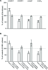
Metabotropic glutamate (Glu) receptors (mGluRs) and GABAB receptors are highly expressed at presynaptic sites. To verify the possibility that the two classes of metabotropic receptors contribute to axon terminals heterogeneity, we studied the localization of mGluR1α, mGluR5, mGluR2/3, mGluR7, and GABAB1 in VGLUT1-, VGLUT2-, and VGAT- positive terminals in the cerebral cortex of adult rats. VGLUT1-positive puncta expressed mGluR1α (∼5%), mGluR5 (∼6%), mGluR2/3 (∼22%), mGluR7 (∼17%), and GABAB1 (∼40%); VGLUT2-positive terminals expressed mGluR1α (∼10%), mGluR5 (∼11%), mGluR2/3 (∼20%), mGluR7 (∼28%), and GABAB1 (∼25%); whereas VGAT-positive puncta expressed mGluR1α (∼27%), mGluR5 (∼24%), mGluR2/3 (∼38%), mGluR7 (∼31%), and GABAB1 (∼19%). Control experiments ruled out the possibility that postsynaptic mGluRs and GABAB1 might have significantly biased our results. We also performed functional assays in synaptosomal preparations, and showed that all agonists modify Glu and GABA levels, which return to baseline upon exposure to antagonists. Overall, these findings indicate that mGluR1α, mGluR5, mGluR2/3, mGluR7, and GABAB1 expression differ significantly between glutamatergic and GABAergic axon terminals, and that the robust expression of heteroreceptors may contribute to the homeostatic regulation of the balance between excitation and inhibition.
Keywords: GABA; autoreceptors; cerebral cortex; glutamate; heteroreceptors; metabotropic receptors.
Figures


Similar articles
-
Presynaptic mGlu1 Receptors Control GABAB Receptors in an Antagonist-Like Manner in Mouse Cortical GABAergic and Glutamatergic Nerve Endings.Front Mol Neurosci. 2018 Sep 18;11:324. doi: 10.3389/fnmol.2018.00324. eCollection 2018. Front Mol Neurosci. 2018. PMID: 30279647 Free PMC article.
-
Heterogeneity of glutamatergic and GABAergic release machinery in cerebral cortex.Neuroscience. 2007 Jun 8;146(4):1829-40. doi: 10.1016/j.neuroscience.2007.02.060. Epub 2007 Apr 19. Neuroscience. 2007. PMID: 17445987
-
High level of mGluR7 in the presynaptic active zones of select populations of GABAergic terminals innervating interneurons in the rat hippocampus.Eur J Neurosci. 2003 Jun;17(12):2503-20. doi: 10.1046/j.1460-9568.2003.02697.x. Eur J Neurosci. 2003. PMID: 12823458
-
GABA(B) and group I metabotropic glutamate receptors in the striatopallidal complex in primates.J Anat. 2000 May;196 ( Pt 4)(Pt 4):555-76. doi: 10.1046/j.1469-7580.2000.19640555.x. J Anat. 2000. PMID: 10923987 Free PMC article. Review.
-
Presynaptic metabotropic glutamate and GABAB receptors.Handb Exp Pharmacol. 2008;(184):373-407. doi: 10.1007/978-3-540-74805-2_12. Handb Exp Pharmacol. 2008. PMID: 18064420 Review.
Cited by
-
Functional Cross-Talk between Adenosine and Metabotropic Glutamate Receptors.Curr Neuropharmacol. 2019;17(5):422-437. doi: 10.2174/1570159X16666180416093717. Curr Neuropharmacol. 2019. PMID: 29663888 Free PMC article. Review.
-
Metabotropic and ionotropic glutamate receptors as potential targets for the treatment of alcohol use disorder.Neurosci Biobehav Rev. 2017 Jun;77:14-31. doi: 10.1016/j.neubiorev.2017.02.024. Epub 2017 Feb 24. Neurosci Biobehav Rev. 2017. PMID: 28242339 Free PMC article. Review.
-
Presynaptic Release-regulating Metabotropic Glutamate Receptors: An Update.Curr Neuropharmacol. 2020;18(7):655-672. doi: 10.2174/1570159X17666191127112339. Curr Neuropharmacol. 2020. PMID: 31775600 Free PMC article. Review.
-
Regulation of Glutamatergic Activity via Bidirectional Activation of Two Select Receptors as a Novel Approach in Antipsychotic Drug Discovery.Int J Mol Sci. 2020 Nov 20;21(22):8811. doi: 10.3390/ijms21228811. Int J Mol Sci. 2020. PMID: 33233865 Free PMC article. Review.
-
Presynaptic mGlu1 Receptors Control GABAB Receptors in an Antagonist-Like Manner in Mouse Cortical GABAergic and Glutamatergic Nerve Endings.Front Mol Neurosci. 2018 Sep 18;11:324. doi: 10.3389/fnmol.2018.00324. eCollection 2018. Front Mol Neurosci. 2018. PMID: 30279647 Free PMC article.
References
-
- Alonso-Nanclares L., Minelli A., Melone M., Edwards R. H., Defelipe J., Conti F. (2004). Perisomatic glutamatergic axon terminals: a novel feature of cortical synaptology revealed by vesicular glutamate transporter 1 immunostaining. Neuroscience 123 547–556. 10.1016/j.neuroscience.2003.09.033 - DOI - PubMed
LinkOut - more resources
Full Text Sources
Other Literature Sources

