. 2015 Sep 3:6:915.
doi: 10.3389/fmicb.2015.00915. eCollection 2015.

Pseudomonas aeruginosa lasI/rhlI quorum sensing genes promote phagocytosis and aquaporin 9 redistribution to the leading and trailing regions in macrophages

Affiliations

Pseudomonas aeruginosa lasI/rhlI quorum sensing genes promote phagocytosis and aquaporin 9 redistribution to the leading and trailing regions in macrophages

Angelika Holm et al. Front Microbiol. .

Abstract

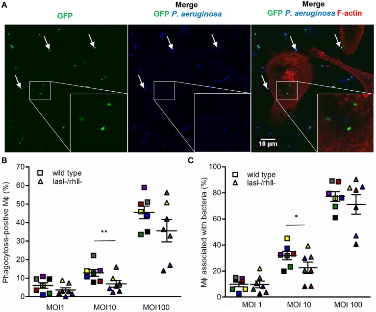
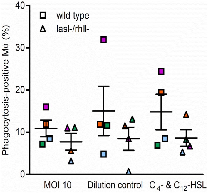
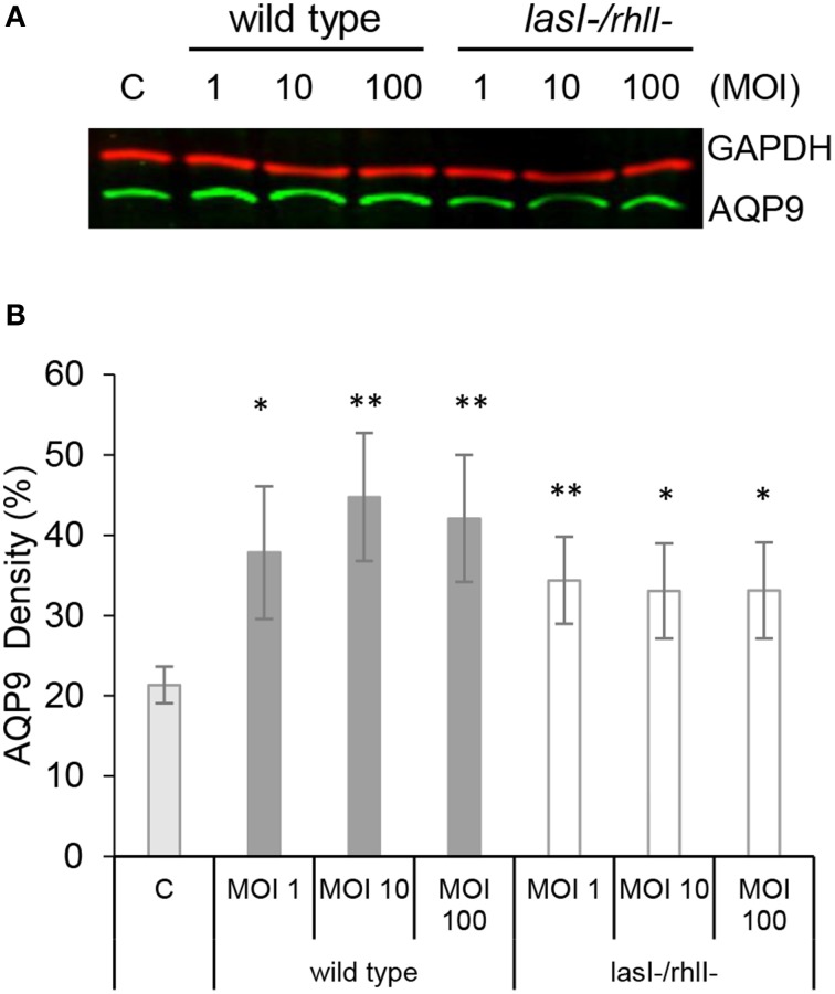
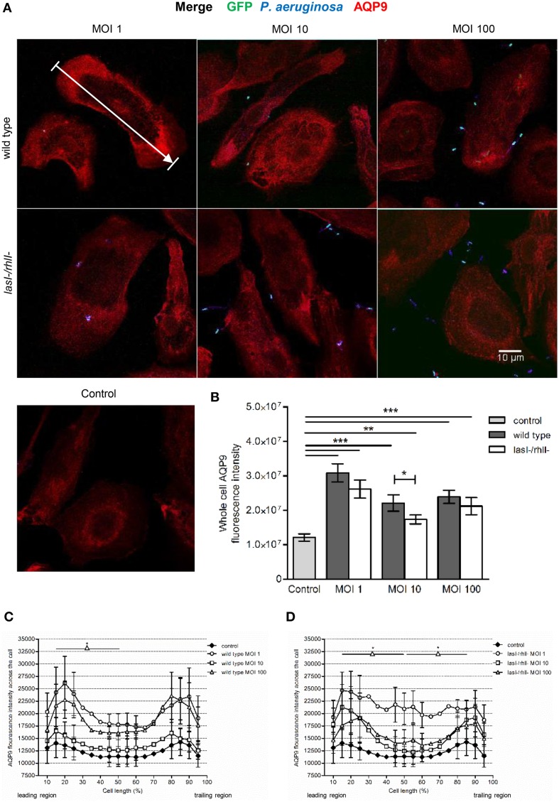
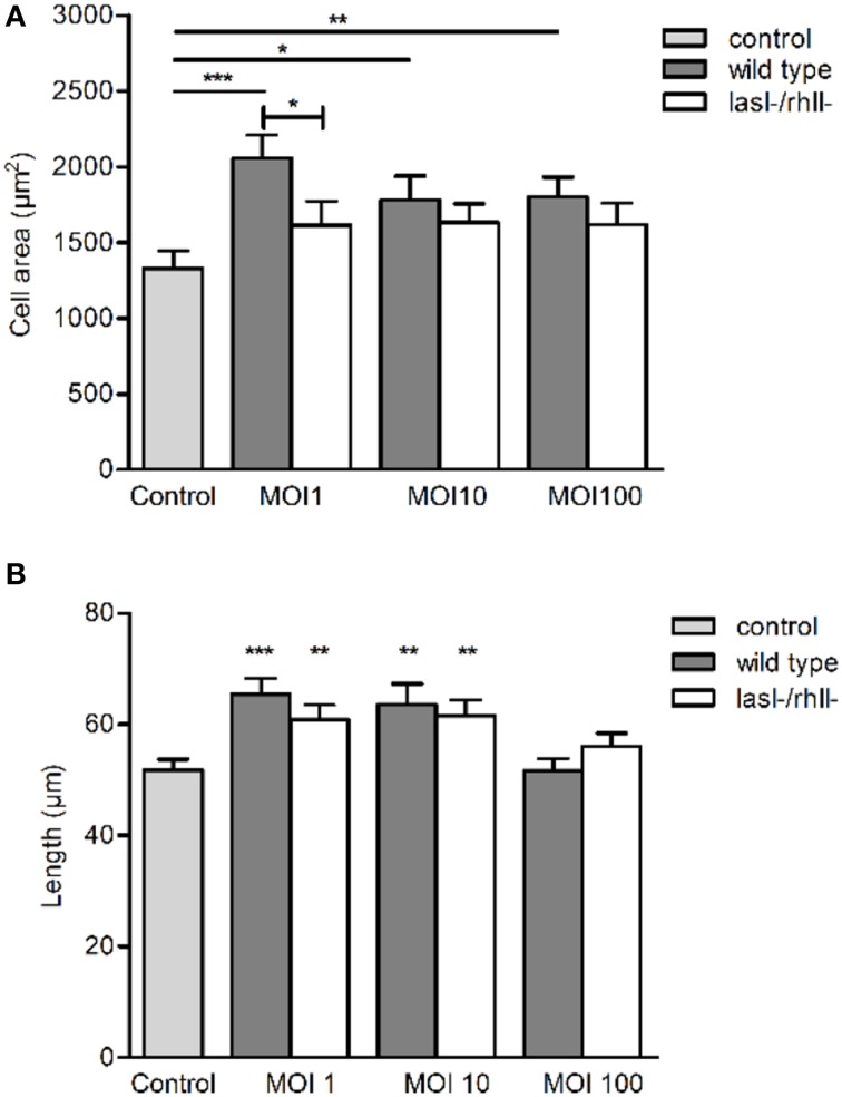
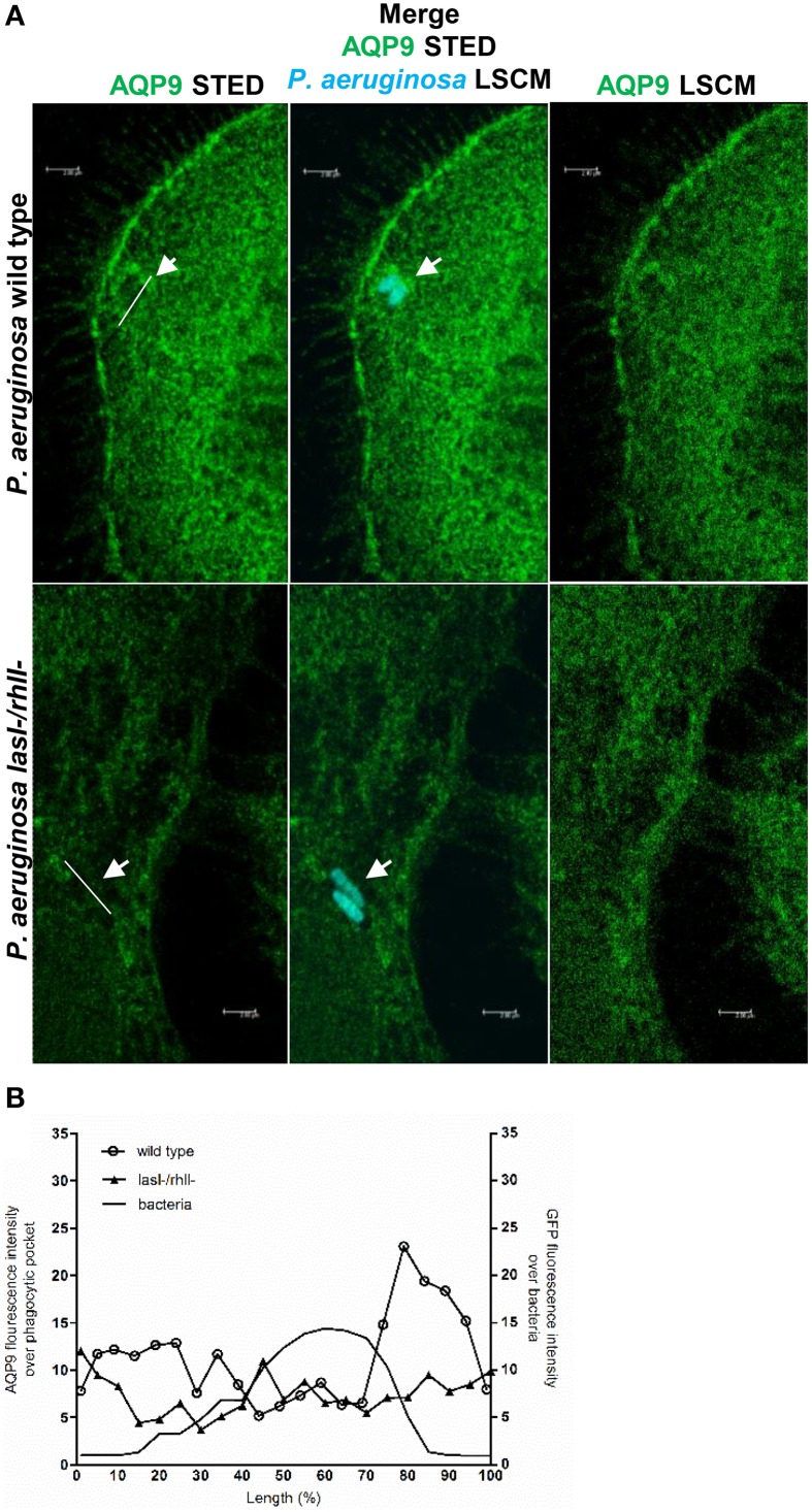
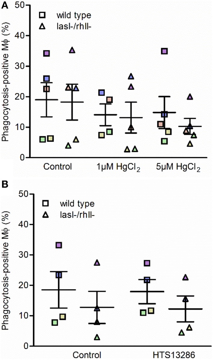
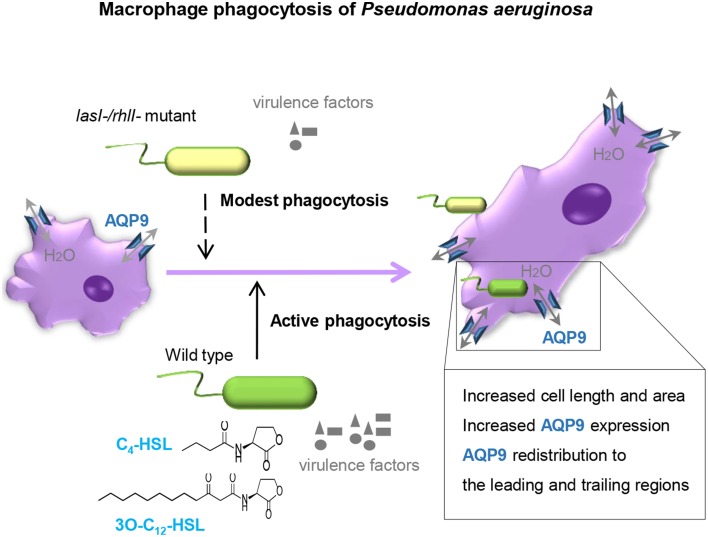
Pseudomonas aeruginosa controls production of its multiple virulence factors and biofilm development via the quorum sensing (QS) system. QS signals also interact with and affect the behavior of eukaryotic cells. Host water homeostasis and aquaporins (AQP) are essential during pathological conditions since they interfere with the cell cytoskeleton and signaling, and hereby affect cell morphology and functions. We investigated the contribution of P. aeruginosa QS genes lasI/rhlI to phagocytosis, cell morphology, AQP9 expression, and distribution in human macrophages, using immunoblotting, confocal, and nanoscale imaging. Wild type P. aeruginosa with a functional QS system was a more attractive prey for macrophages than the lasI/rhlI mutant lacking the production of QS molecules, 3O-C12-HSL, and C4-HSL, and associated virulence factors. The P. aeruginosa infections resulted in elevated AQP9 expression and relocalization to the leading and trailing regions in macrophages, increased cell area and length; bacteria with a functional QS system lasI/rhlI achieved stronger responses. We present evidence for a new role of water fluxes via AQP9 during bacteria-macrophage interaction and for the QS system as an important stimulus in this process. These novel events in the interplay between P. aeruginosa and macrophages may influence on the outcome of infection, inflammation, and development of disease.

Keywords: N-acylhomoserine lactone; aquaporin; host-bacteria relationship; innate immunity; macrophage; quorum sensing; water homeostasis.


Figures

Figure 1
Figure 1
Binding and phagocytosis of wild type P. aeruginosa and its lasI-/rhlI- mutant. (A) Macrophages were infected with GFP (green) wild type bacteria or lasI-/rhlI- mutant, stained for P. aeruginosa (blue) and F-actin (red), and analyzed by LSCM. White squares show three ingested bacteria distinguished by sole GFP (green). White arrows point to bound bacteria recognized by combined GFP (green) and P. aeruginosa (blue). Bar 10 μm. (B) Quantification of phagocytosis presented as the percentage of phagocytic-positive (containing ingested bacteria) cells among total macrophages. (C) Quantification of binding presented as the percentage of cells containing bound and ingested bacteria (associated bacteria) among total macrophages. Shown are the mean ± SE of seven independent experiments performed at separate days from different donors (color coded). The means ± SE are based on 100–200 cells for each condition per experiment. Significant differences were considered at *P < 0.05 and **P < 0.01, as calculated by two-tailed paired Student's t-test.
Figure 2
Figure 2
Functional complementation of phagocytosis with AHL. Macrophages were pretreated with 25 μM C4-HSL and 50 μM 3O-C12-HSL (C4- C12-HSL), or 0.02% DMSO as a vehicle control (Dilution control), or not-pretreated (MOI 10) for 4 h before 1-h infection with P. aeruginosa wild type or lasI-/rhlI- mutant, at MOI 10. Quantification of phagocytosis presented as the percentage of phagocytic-positive (containing ingested bacteria) cells among total macrophages. Shown are mean ± SE of four independent experiments performed at separate days from four different donors (color coded). The means ± SE are based on 100–200 cells for each condition per experiment.
Figure 3
Figure 3
AQP9 protein levels are increased in macrophages infected with P. aeruginosa. (A) Macrophages were either non-infected (C) or infected with wild type P. aeruginosa or lasI-/rhlI- mutant at MOI 1, 10, and 100 for 1 h. Total cellular protein extracts were analyzed with Western blot for AQP9, 31 kDa (lower, green bands) and GAPDH, 36 kDa as a loading control (upper, red bands). The blots are from one representative of four independent experiments. (B) Densitometric analysis. Values are mean ± SE percentage of AQP9 density relative to the loading GAPDH control from four independent experiment performed at separate days from four different donors. Significant differences are indicated when *P < 0.05 and **P < 0.01, as analyzed by Student's t-test.
Figure 4
Figure 4
Effects of P. aeruginosa on the cellular distribution of AQP9. (A) Macrophages were infected with GFP (green) wild type P. aeruginosa, the lasI-/rhlI- mutant, or non-infected (Control), stained for P. aeruginosa (blue), AQP9 (red) and analyzed by LSCM. The data are from one of at least three independent experiments. Bar 10 μm. (B) Quantification of AQP9 immunofluorescence intensity measured as a total integrated intensity of whole macrophage area, of at least 30 cells for each condition. Data are from at least three experiments performed at separate days from different donors. Columns represent means ± SE. Significant differences are indicated when **P < 0.01 and ***P < 0.0001, as analyzed by two-tailed unpaired Student's t-test. (C,D) Quantification of AQP9 immunofluorescence intensity profile measured across the cells in the direction of polarization as indicated by white arrow in (A). Significant differences are indicated when *P < 0.05, as analyzed by one-tailed unpaired Student's t-test.
Figure 5
Figure 5
P. aeruginosa affects cell morphology in macrophages. Macrophages were infected with wild type P. aeruginosa, the lasI-/rhlI- mutant or were non-infected (Control), stained, and imaged as in Figure 4A. (A) Quantification of the macrophage cell area. (B) Quantification of approximated macrophage length (as indicated by white arrow in Figure 4A in the direction of polarization). Columns represent means ± SE of three different experiments with three donors. Significant differences are indicated when *P < 0.05, **P < 0.01 and ***P < 0.0001, as analyzed by two-tailed unpaired Student's t-test.
Figure 6
Figure 6
Nanoscale visualization of AQP9 in macrophages during P. aeruginosa infections. (A) Macrophages were infected with GFP-labeled (cyan) wild type P. aeruginosa or the lasI-/rhlI- mutant or non-infected (not shown). Cells were stained for non-ingested P. aeruginosa (not shown), and AQP9 (green), and analyzed by STED. Bacteria shown at these images were completely ingested (cyan). White arrows points to pocket-like AQP9 accumulation around completely ingested wild type bacteria. Bar 2 μm. (B) Mean AQP9 intensity profiles over phagocytic pockets and mean GFP intensity profile over bacteria from representative images (A) measured as indicated by the white lines.
Figure 7
Figure 7
Effect of water fluxes inhibitors on phagocytosis of P. aeruginosa by macrophages. Macrophages were pretreated with (A) 1 or 5 μM HgCl2, or PBS as a control; (B) 25 μM HTS13286 or 0.25% DMSO as vehicle control for 20 min. Wild type P. aeruginosa or the lasI-/rhlI- mutant at MOI 10 were added for 1 h of infection and phagocytic activity of macrophages were evaluated. Shown are the mean ± SE of the percentage of phagocytosis-positive macrophages. Data are from individual independent experiments performed at separate days from 4 to 5 different donors (color coded).
Figure 8
Figure 8
Proposed model. P. aeruginosa lasI/rhlI quorum sensing genes promote phagocytosis and AQP9 redistribution to the leading and trailing regions in macrophages. The phagocytosis of P. aeruginosa by macrophages seems more effective when QS genes lasI and rhlI responsible for synthesis of two QS molequles 3O-C12-HSL and C4-HSL and associated virulence factors are fully functional. Moreover, the P. aeruginosa infections results in elevated AQP9 expression and relocalization to the leading and trailing regions in macrophages, increased cell area, and length; here, bacteria with a functional QS system lasI/rhlI achieved stronger responses.

Similar articles

Cited by

References

    1. Allen L., Dockrell D. H., Pattery T., Lee D. G., Cornelis P., Hellewell P. G., et al. . (2005). Pyocyanin production by Pseudomonas aeruginosa induces neutrophil apoptosis and impairs neutrophil-mediated host defenses in vivo. J. Immunol. 174, 3643–3649. 10.4049/jimmunol.174.6.3643 - DOI - PubMed
    1. Benga G. (2012). On the definition, nomenclature and classification of water channel proteins (aquaporins and relatives). Mol. Aspects Med. 33, 514–517. 10.1016/j.mam.2012.04.003 - DOI - PubMed
    1. Blom H., Brismar H. (2014). STED microscopy: increased resolution for medical research? J. Intern. Med. 276, 560–578. 10.1111/joim.12278 - DOI - PubMed
    1. Brint J. M., Ohman D. E. (1995). Synthesis of multiple exoproducts in Pseudomonas aeruginosa is under the control of RhlR-RhlI, another set of regulators in strain PAO1 with homology to the autoinducer-responsive LuxR-LuxI family. J. Bacteriol. 177, 7155–7163. - PMC - PubMed
    1. Bylund J., Burgess L. A., Cescutti P., Ernst R. K., Speert D. P. (2006). Exopolysaccharides from Burkholderia cenocepacia inhibit neutrophil chemotaxis and scavenge reactive oxygen species. J. Biol. Chem. 281, 2526–2532. 10.1074/jbc.M510692200 - DOI - PubMed