. 2015 Nov;11(11):870-7.
doi: 10.1038/nchembio.1926. Epub 2015 Sep 21.

Translocation-coupled DNA cleavage by the Type ISP restriction-modification enzymes

Affiliations

Translocation-coupled DNA cleavage by the Type ISP restriction-modification enzymes

Mahesh K Chand et al. Nat Chem Biol. 2015 Nov.

Abstract

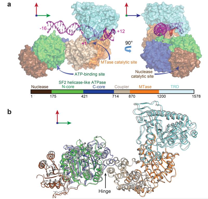
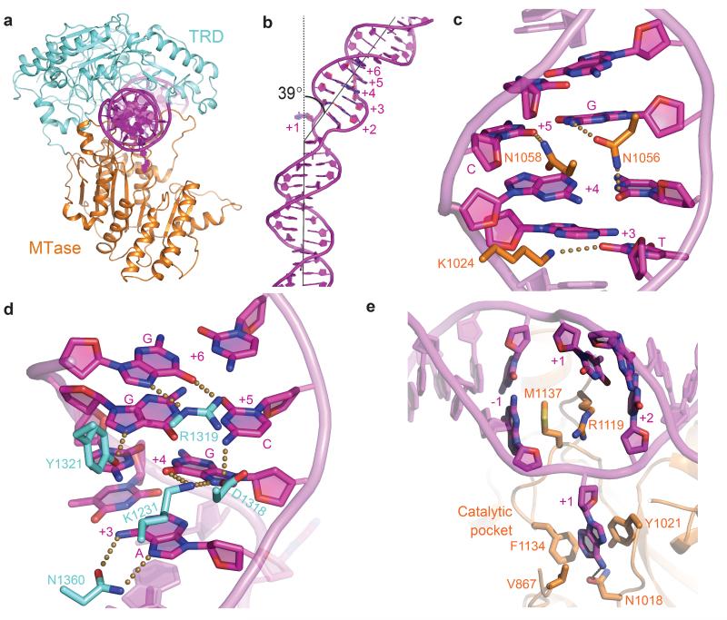
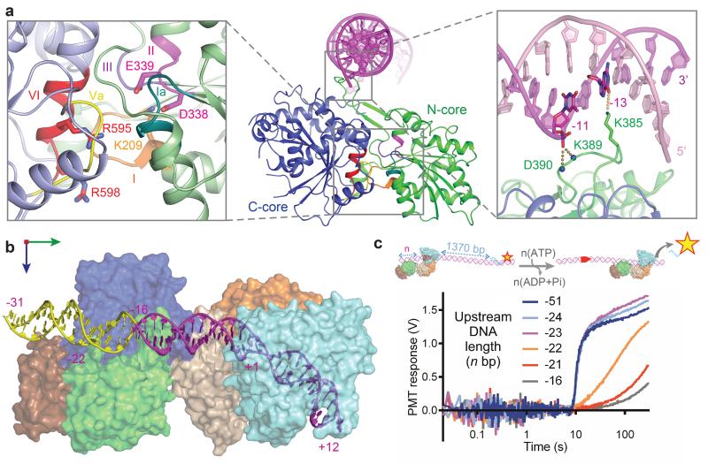
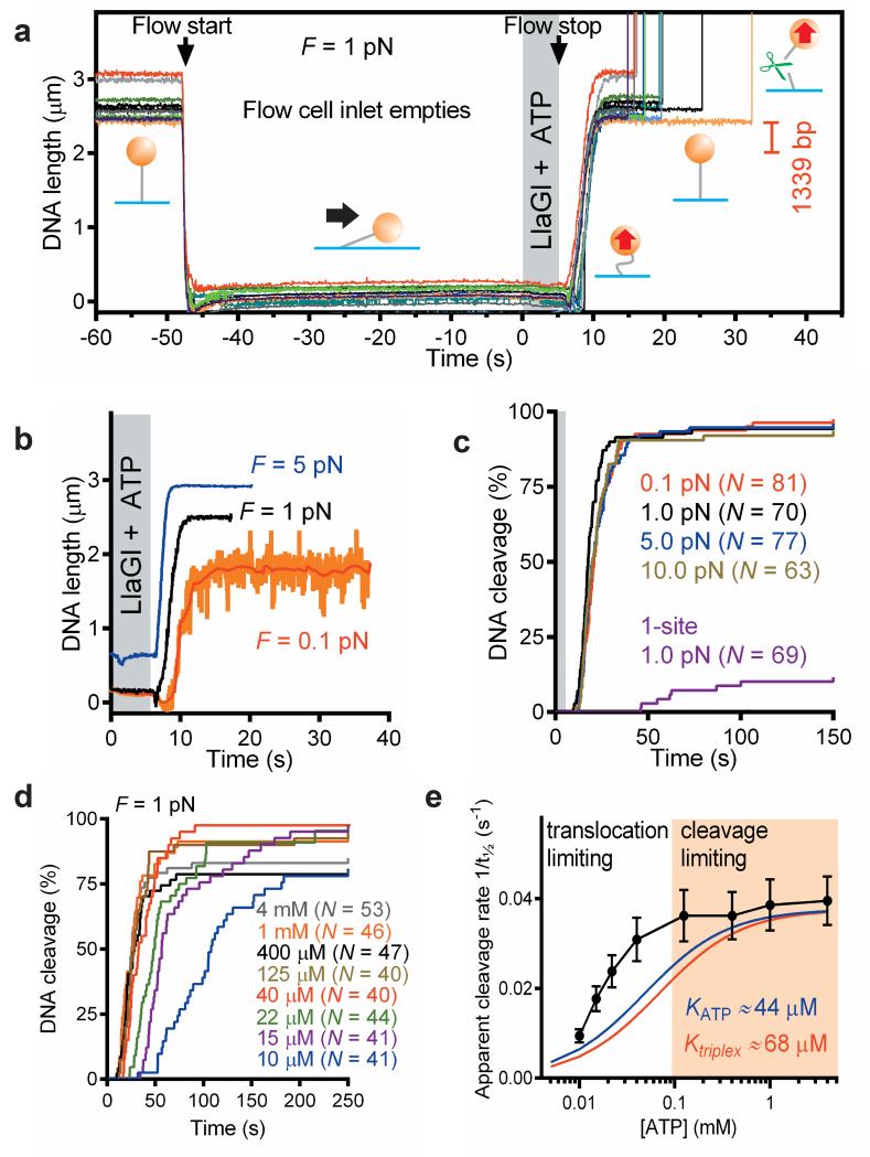
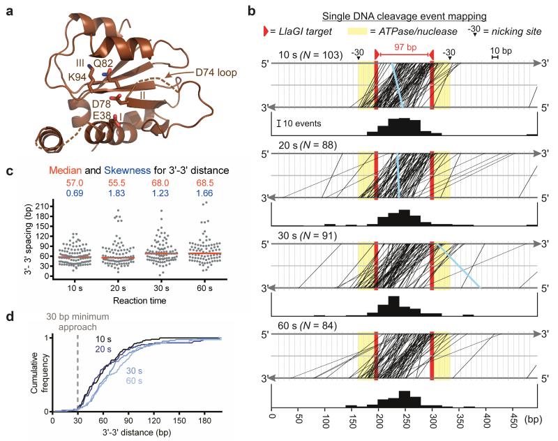
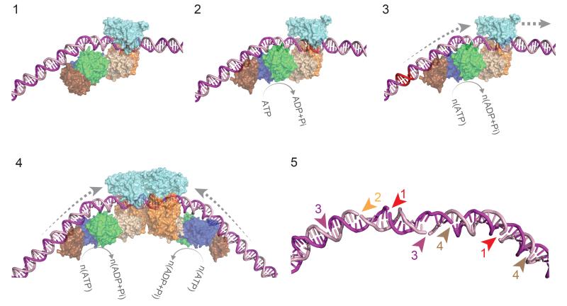
Production of endonucleolytic double-strand DNA breaks requires separate strand cleavage events. Although catalytic mechanisms for simple, dimeric endonucleases are known, there are many complex nuclease machines that are poorly understood. Here we studied the single polypeptide Type ISP restriction-modification (RM) enzymes, which cleave random DNA between distant target sites when two enzymes collide after convergent ATP-driven translocation. We report the 2.7-Å resolution X-ray crystal structure of a Type ISP enzyme-DNA complex, revealing that both the helicase-like ATPase and nuclease are located upstream of the direction of translocation, an observation inconsistent with simple nuclease-domain dimerization. Using single-molecule and biochemical techniques, we demonstrate that each ATPase remodels its DNA-protein complex and translocates along DNA without looping it, leading to a collision complex in which the nuclease domains are distal. Sequencing of the products of single cleavage events suggests a previously undescribed endonuclease model, where multiple, stochastic strand-nicking events combine to produce DNA scission.


Figures

Figure 1
Figure 1. Architecture of the modular Type ISP RM enzymes
a, Surface representation of LlaBIII-DNA. b, Superposition of the two chains of LlaBIII reveals conformational plasticity. The coupler domain (residues 715 to 870) of Chain B (grey) was superimposed onto the coupler domain of chain A using the Secondary-structure matching method in Coot. The structural domains of Chain A are coloured as in Fig. 1b. The hinge about which the nuclease-ATPase domains move with respect to the coupler is indicated (Supplementary Table 1).
Figure 2
Figure 2. DNA target recognition by LlaBIII
a, Ribbon diagram of the MTase-TRD clamp encircling the target. b, Extent of DNA bending. c, Minor groove readout by MTase. d, Major groove readout by TRD. e, MTase-DNA contacts stabilising the flipped adenine. Dotted lines represent interactions within hydrogen bonding distance (less than or equal to 3.5 Å).
Figure 3
Figure 3. Architecture and upstream positioning of the ATPase domains
a, ATPase architecture highlighting: (left inset) conserved SF2 helicase-like motifs, with ATP-interacting residues as sticks; (Right inset) DNA interaction of loop S383-D390, with side-chain of K385 as sticks and main chain nitrogens of K389 and D390 as spheres. b, Model of nuclease-ATPase interactions with an extended DNA (in yellow). c, The effect of upstream DNA length on triplex displacement. (cartoon) LlaGI motor activity initiated with ATP was monitored on DNA with varying upstream DNA.
Figure 4
Figure 4. Loop-independent translocation
a, DNA cleavage events (upward spikes) following transient introduction of LlaGI and ATP into the flow cell. DNA length change expected of looping is indicated. Lines are raw data (60 Hz). Flow is started as indicated to empty the inlet. Bead positions change because of flow and cell distortion due to hydrodynamic pressure. The LlaGI + ATP solution is added and the flow stopped after ~5 s. The DNA relaxes back to full extension slowly because of hydrostatic re-equilibration of the cell. b, examples of loop-independent cleavage at a range of stretching forces. Lines are raw data (31 Hz) except the red line (1 s filter). c, Cumulative dsDNA cleavage for N events on head-to-head DNA at 0.1-10 pN and on a one-site DNA (Supplementary Fig. 12) at 1.0 pN. d, Cumulative dsDNA cleavage for N events on head-to-head DNA at 1 pN at a range of ATP concentrations. e, ATP dependence of the apparent cleavage rate (taken from t½, the time to reach 50% cleavage in f). Blue and red lines are simulations of a hyperbolic relationship for the previously-determined apparent KM,app values for ATP hydrolysis (KATP) and DNA translocation (Ktriplex). Error bars were calculated from the average values divided by the square root of number of points (N) in the bin.
Figure 5
Figure 5. Single cleavage event mapping
a, Mrr-family nuclease, highlighting catalytic motifs and residues. b, DNA cleavage by LlaGI at each time point is plotted as top/bottom strand locations joined by a line (rare 5′-5′ overhangs in blue). Bar graphs are the collision centroids (20 bp bins). c, Scatter plot of the outermost 3′-3′ cleavage distances, with median (red line) and skewness values (the latter showing a time-dependent increase in data asymmetry). d, Cumulative frequency (normalised for N events) of outermost 3′-3′ cleavage distances.
Figure 6
Figure 6. Model for loop-independent DNA translocation and extensive nucleolytic DNA processing
Model for loop-independent DNA translocation and extensive nucleolytic DNA processing. (1) The pre-initiation complex. The nuclease is in an inactive conformation. (2) The ATPase cycle loosens the MTase-TRD grip on the DNA. (3) dsDNA translocation downstream of the target (red). (4) Convergence of two enzymes initially brings the nucleases approximately 75 bp apart. (5) Example of stochastic “DNA shredding” by a collision complex. Numbers are the order of the nicking events.

References

Reference list

    1. Dryden DTF, Murray N, Rao DN. Nucleoside triphosphate-dependent restriction enzymes. Nucleic Acids Res. 2001;29:3728–3741. - PMC - PubMed
    1. Labrie SJ, Samson JE, Moineau S. Bacteriophage resistance mechanisms. Nature Rev. Microbiol. 2010;8:317–327. - PubMed
    1. Linn S, Arber W. Host specificity of DNA produced by Escherichia coli, X. In vitro restriction of phage fd replicative form. Proc. Natl. Acad. Sci. USA. 1968;59:1300–1306. - PMC - PubMed
    1. Meselson M, Yuan R. DNA restriction enzyme from E. coli. Nature. 1968;217:1110–1114. - PubMed
    1. Loenen WAM, Dryden DTF, Raleigh EA, Wilson GG. Type I restriction enzymes and their relatives. Nucleic Acids Res. 2014;42:20–44. - PMC - PubMed

Methods-only references

    1. LeMaster DM, Richards FM. NMR sequential assignment of Escherichia coli thioredoxin utilizing random fractional deuteriation. Biochemistry. 1988;27:142–150. - PubMed
    1. Kabsch W. XDS. Acta Crystallogr. D Biol.Crystallogr. 2010;66:125–132. - PMC - PubMed
    1. French S, Wilson K. On the treatment of negative intensity observations. Acta Crystallogr. A. 1978;34:517–525.
    1. Read RJ, McCoy AJ. Using SAD data in Phaser. Acta Crystallogr. D Biol.Crystallogr. 2011;67:338–344. - PMC - PubMed
    1. Cowtan KD, Zhang KYJ. Density modification for macromolecular phase improvement. Prog. Biophy. Mol. Biol. 1999;72:245–270. - PubMed

Publication types

MeSH terms

Associated data

LinkOut - more resources