. 2015 Nov;18(11):1679-1686.
doi: 10.1038/nn.4119. Epub 2015 Sep 21.

Hierarchical nesting of slow oscillations, spindles and ripples in the human hippocampus during sleep

Affiliations

Hierarchical nesting of slow oscillations, spindles and ripples in the human hippocampus during sleep

Bernhard P Staresina et al. Nat Neurosci. 2015 Nov.

Abstract

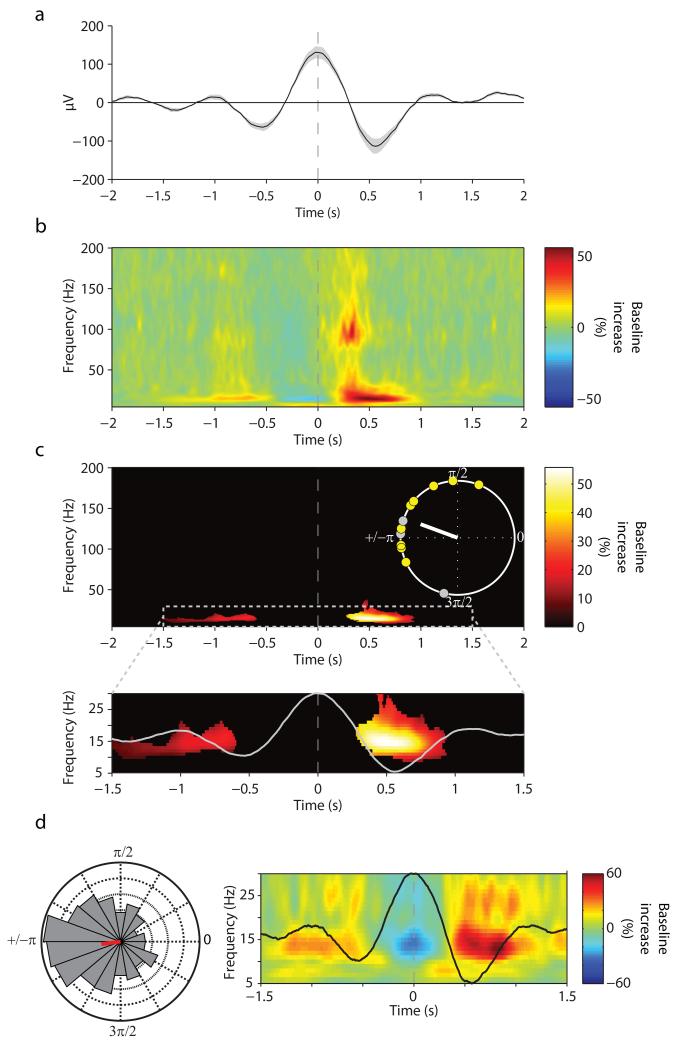
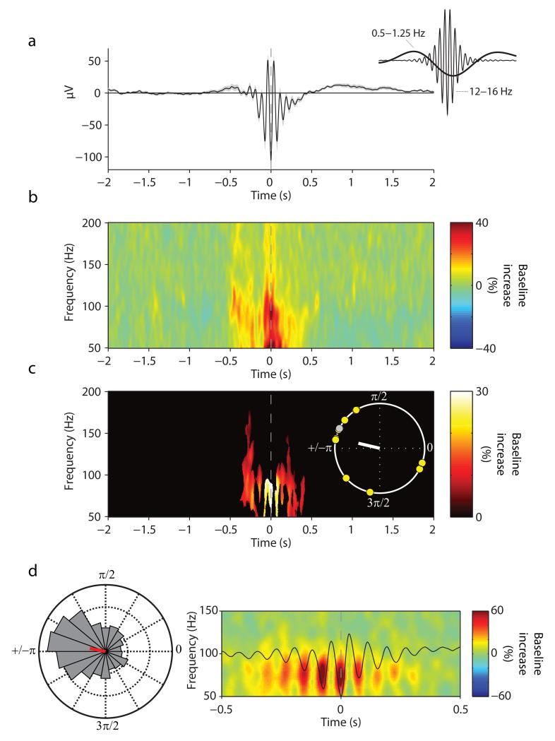
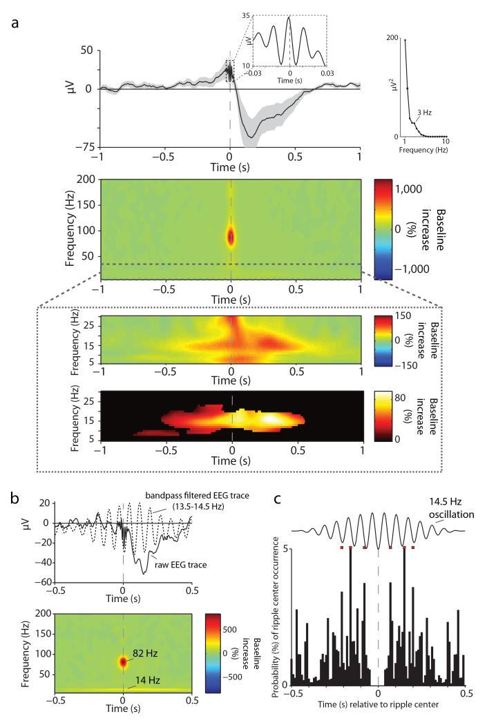
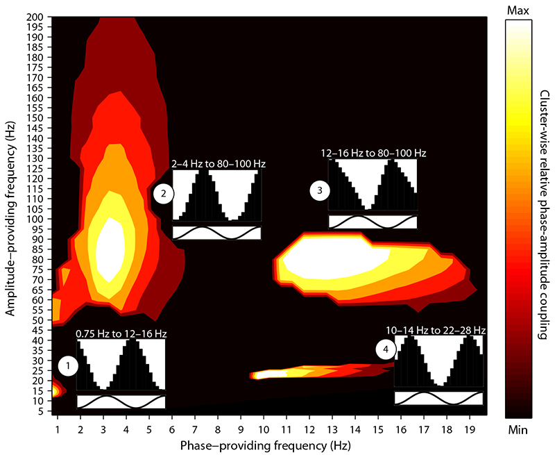
During systems-level consolidation, mnemonic representations initially reliant on the hippocampus are thought to migrate to neocortical sites for more permanent storage, with an eminent role of sleep for facilitating this information transfer. Mechanistically, consolidation processes have been hypothesized to rely on systematic interactions between the three cardinal neuronal oscillations characterizing non-rapid eye movement (NREM) sleep. Under global control of de- and hyperpolarizing slow oscillations (SOs), sleep spindles may cluster hippocampal ripples for a precisely timed transfer of local information to the neocortex. We used direct intracranial electroencephalogram recordings from human epilepsy patients during natural sleep to test the assumption that SOs, spindles and ripples are functionally coupled in the hippocampus. Employing cross-frequency phase-amplitude coupling analyses, we found that spindles were modulated by the up-state of SOs. Notably, spindles were found to in turn cluster ripples in their troughs, providing fine-tuned temporal frames for the hypothesized transfer of hippocampal memory traces.


Figures

Fig. 1
Fig. 1. Implantation and sleep recordings
a. Post-operative MRI (top) and co-registered pre-operative MRI (bottom) for a sample participant, showing a depth electrode implanted along the longitudinal axis of the hippocampus (Supplementary Fig. 1 shows electrode locations for all participants). Cross-hair is placed on the electrode taken forward to the group analysis (Experimental Procedures). b. Graphic depiction of time spent in different sleep stages across one recording night (hypnogram) for a sample participant.
Fig. 2
Fig. 2
Event-locked analysis of hippocampal SO-spindle PAC. (a) Grand average unfiltered iEEG trace across participants (mean ± s.e.m.), aligned to the maximum of the SO down-state (peak, time 0). (b) Average of SO down-state-locked TFR (% change from pre-event baseline). Y-axis starts at 5 Hz to circumvent the dominance of power in the SO range. (c) Statistically significant change from pre-event baseline (P < .05, corrected). Inset shows unit circle of preferred phases of the SO-spindle modulation for each participant, which illustrates the preferred clustering of spindle power towards the SO up-state (160°, white line). Yellow circles represent participants whose Rayleigh test for non-uniformity was significant at P < .05. (d) Data from a sample participant. (right) average SO and TFR, zoomed in on −1.5 s to +1.5 s and on 5–30 Hz to highlight the nesting of spindle power in the SO up-state. (left) normalized histogram (18 bins, 20° each) of preferred SO-spindle modulation phases across all detected SO events (N=1015), resulting in an average preferred phase of 187° for this participant (red line).
Fig. 3
Fig. 3
Event-locked analysis of hippocampal spindle-ripple PAC. (a) Grand average unfiltered iEEG trace across participants (mean ± s.e.m.), aligned to the maximum of the spindle trough (time 0). Note that the spindle’s mean potential is below zero (−250 ms to +250 ms, t(11) = 3.94, P = .002), reflecting the grouping of spindles in the SO trough (as shown in Fig. 2). This is further illustrated in the inset, which shows the grand average iEEG trace bandpass filtered from 0.5–1.25 Hz (SO range) and from 12–16 Hz (spindle range), respectively. (b) Average of spindle-trough-locked TFR (% change from pre-event baseline). Y-axis starts at 50 Hz to circumvent the dominance of power in the spindle range. (c) Statistically significant change from pre-event baseline (P < .05, corrected). Inset shows unit circle of preferred phases of the spindle-ripple modulation for each participant, which illustrates the preferred clustering of ripple power in the spindle trough (167°, white line). Yellow circles represent participants whose Rayleigh test for non-uniformity was significant at P < .05. (d) Data from one participant. (right) average spindle and TFR, zoomed in on −0.5 s to 0.5 s and on 50–150 Hz to illustrate the nesting of ripple power within spindle troughs. (left) normalized histogram (18 bins, 20° each) of preferred spindle-ripple modulation phases across all detected spindle events (N=1487), resulting in an average preferred phase of 169° for this participant (red line).
Fig. 4
Fig. 4
Hippocampal ripples. (a) (top) Grand average unfiltered iEEG trace across participants (mean ± s.e.m.), aligned to the maximum of the ripple peak (time 0). The first inset highlights the oscillatory nature of the detected events in the unfiltered raw EEG. The second inset shows the result from a spectral analysis of the grand average EEG (calculated from −1 s to +1 s via FieldTrip’s ‘mtmfft’ function for frequencies from 1–10 Hz in steps of .5 Hz), revealing a spectral peak at 3 Hz (delta wave). (middle) Average of ripple-peak-locked TFR (% change from pre-event baseline), highlighting the band-limited nature of ripples. (bottom) Zoom into 5–30 Hz, showing the grand-average TFR before (top) and after (bottom) statistical thresholding (P < .05, corrected) to highlight the ripple-locked increase in spindle power. (b) Data from a sample participant. (top) ripple-locked average raw EEG trace (solid line) with the bandpass filtered trace (13.5–14.5 Hz; dotted line) superimposed to highlight the nesting of ripples in spindle troughs. (bottom). Corresponding ripple-locked TFR, highlighting that participant’s maxima in the ripple- and spindle ranges. (c) Ripple-ripple peri-event time histogram (PETH). Occurrence probabilities of other ripples relative to individual ripples’ center time. A synthetic 14.5 Hz oscillation is superimposed to illustrate the periodicity of ripple occurrences following successive spindle cycles. Red squares project the histogram maxima from −250 ms to +250 ms relative to the ripple center to better visualize the correspondence of occurrences with individual spindle cycles.
Fig. 5
Fig. 5
Hippocampal phase-amplitude coupling (PAC) during NREM sleep. Clusters showing a significant Modulation Index (MI) when comparing (i) NREM PAC vs. WAKE PAC and (ii) NREM PAC vs. trial-shuffled NREM surrogate data. Both contrasts were thresholded at P < .05 (two-tailed paired-sample t-tests), and significant clusters were defined as a minimum of 25 contiguous frequency pairs showing significant differences in both contrasts. Effect sizes are scaled within each cluster to a maximum of 1 and a minimum of 0 (separate unscaled Comodulograms for each comparison are shown in Supplementary Fig. 9). Insets show average amplitudes of the modulated frequency as a function of 1.5 cycles of the modulating frequency’s phase, with y-axes ranging from minimum to maximum amplitude modulation.

References

    1. Born J, Rasch B, Gais S. Sleep to remember. The Neuroscientist. 2006;12:410–424. - PubMed
    1. Buzsáki G. The hippocampo-neocortical dialogue. Cerebral Cortex. 1996;6:81–92. - PubMed
    1. McClelland JL, McNaughton BL, O’Reilly RC. Why there are complementary learning systems in the hippocampus and neocortex: insights from the successes and failures of connectionist models of learning and memory. Psychological review. 1995;102:419. - PubMed
    1. Rasch B, Born J. About sleep’s role in memory. Physiological reviews. 2013;93:681–766. - PMC - PubMed
    1. Diekelmann S, Born J. The memory function of sleep. Nature Reviews Neuroscience. 2010;11:114–126. - PubMed

Methods references

    1. Iber C, Ancoli-Israel S, Chesson A, Quan SF. The AASM manual for the scoring of sleep and associated events: rules, terminology and technical specifications. 2007.
    1. Oostenveld R, Fries P, Maris E, Schoffelen J-M. FieldTrip: Open Source Software for Advanced Analysis of MEG, EEG, and Invasive Electrophysiological Data. Computational Intelligence and Neuroscience. 2011 - PMC - PubMed
    1. Berens P. CircStat: a Matlab Toolbox for Circular Statistics. Journal of Statistical Software. 2009;31
    1. Maris E, Oostenveld R. Nonparametric statistical testing of EEG-and MEG-data. Journal of neuroscience methods. 2007;164:177–190. - PubMed
    1. Mormann F, et al. Phase/amplitude reset and theta–gamma interaction in the human medial temporal lobe during a continuous word recognition memory task. Hippocampus. 2005;15:890–900. - PubMed

Publication types