Review
. 2015 Sep 15;16(9):22368-401.
doi: 10.3390/ijms160922368.

Neuroprotective Strategies after Neonatal Hypoxic Ischemic Encephalopathy

Affiliations
Review

Neuroprotective Strategies after Neonatal Hypoxic Ischemic Encephalopathy

Brandon J Dixon et al. Int J Mol Sci. .

Abstract

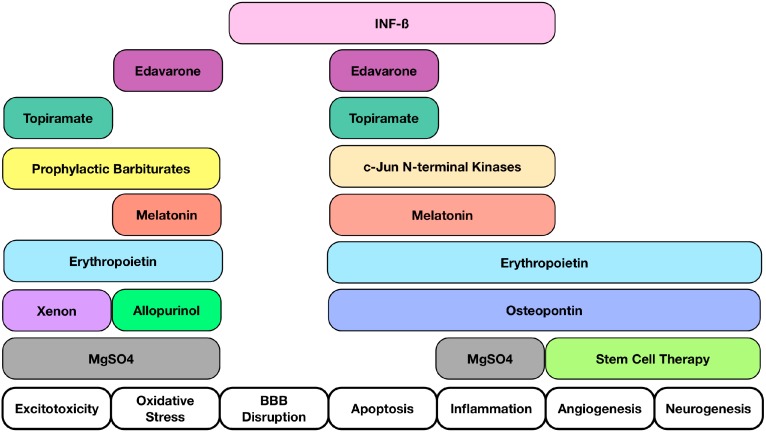
Neonatal hypoxic ischemic encephalopathy (HIE) is a devastating disease that primarily causes neuronal and white matter injury and is among the leading cause of death among infants. Currently there are no well-established treatments; thus, it is important to understand the pathophysiology of the disease and elucidate complications that are creating a gap between basic science and clinical translation. In the development of neuroprotective strategies and translation of experimental results in HIE, there are many limitations and challenges to master based on an appropriate study design, drug delivery properties, dosage, and use in neonates. We will identify understudied targets after HIE, as well as neuroprotective molecules that bring hope to future treatments such as melatonin, topiramate, xenon, interferon-beta, stem cell transplantation. This review will also discuss some of the most recent trials being conducted in the clinical setting and evaluate what directions are needed in the future.

Keywords: intervention strategy; neonatal hypoxic ischemic encephalopathy; neuroprotection; therapeutic strategy.


Figures

Figure 1
Figure 1
Interventional Targets Following Hypoxic Ischemic Encephalopathy (HIE). A summary of potential interventional targets following HIE along with molecules that can exert multiple properties. Interventional targets consist of Excitotoxicity, Oxidative Stress, Blood Brain Barrier Disruption, Apoptosis, Inflammation, Angiogenesis, and Neurogenesis. Multiple target sites suggest that a multi-targeted approach is beneficial after HIE.
Figure 2
Figure 2
(a) Vasculature Changes and Primary Energy Failure (Phase I) Legend: A visual representation of the first phase of HIE. Detrimental changes to the vasculature following an HIE insult lead to loss of autoregulation and severe lowering of the systemic arterial blood pressure. This causes a decrease in oxygen, depletion of ATP, as well as increases in excitotoxicity, intracellular calcium, oxidative stress, and mitochondrial dysfunction; (b) Secondary Energy Failure (Phase II). Legend: A schematic representation of the second phase of HIE reveals continued excitotoxicity, oxidative stress, and mitochondrial dysfunction; (c) Chronic Inflammation (Phase III). A pictorial representation of the third phase of HIE shows injury to microglia, neurons, and astrocytes leads to continuous release of cytokines and other detrimental factors causing chronic inflammation which in turn leads to epigenetic changes, as well as impairments of synaptogenesis, axonal growth, and neurogenesis.
Figure 3
Figure 3
Stem Cell Transplantation in Animal Models of HIE. Summary of stem cell transplantation studies in various animal models of HIE. Human Dental Pulp Stem Cells (DPSCs); Hypoxic Ischemic Encephalopathy (HIE); Mononuclear Cells (MNCs); Mesenchymal Stem Cells (MSCs); Neuronal Stem Cells (NSC); Based upon cell dose, cell type, transplantation timing, and administration route.
Figure 4
Figure 4
Proposed Mechanisms of Current Clinical Trials. Cord blood infusions are rich with hematopoietic stem cells and neurotrophic factors that have numerous effects such as immunomodulation, reduction of microglia and T-lymphocyte infiltration, as well as the potential to increase neurogenesis and an angiogenesis. It is believed that topiramate is able to block sodium channels and high voltage-activated calcium currents after HIE. Xenon is believed to bind at the glycine site of the NMDA receptor and inhibit its downstream effects. Similarly magnesium sulfate also inhibits the NMDA receptor by binding to the magnesium site of the receptor. Allopurinol is predicted to provide neuroprotection by directly scavenging hydroxyl radicals after HIE injury.

References

    1. Shetty J. Neonatal seizures in hypoxic-ischaemic encephalopathy-risks and benefits of anticonvulsant therapy. Dev. Med. Child. Neurol. 2015;57:40–43. doi: 10.1111/dmcn.12724. - DOI - PubMed
    1. Volpe J.J. Perinatal brain injury: From pathogenesis to neuroprotection. Ment. Retard. Dev. Disabil. Res. Rev. 2001;7:56–64. doi: 10.1002/1098-2779(200102)7:1<56::AID-MRDD1008>3.0.CO;2-A. - DOI - PubMed
    1. Doycheva D., Shih G., Chen H., Applegate R., Zhang J.H., Tang J. Granulocyte-colony stimulating factor in combination with stem cell factor confers greater neuroprotection after hypoxic-ischemic brain damage in the neonatal rats than a solitary treatment. Transl. Stroke Res. 2013;4:171–178. doi: 10.1007/s12975-012-0225-2. - DOI - PMC - PubMed
    1. Northington F.J., Chavez-Valdez R., Martin L.J. Neuronal cell death in neonatal hypoxia-ischemia. Ann. Neurol. 2011;69:743–758. doi: 10.1002/ana.22419. - DOI - PMC - PubMed
    1. Gill M.B., Perez-Polo J.R. Hypoxia ischemia-mediated cell death in neonatal rat brain. Neurochem. Res. 2008;33:2379–2389. doi: 10.1007/s11064-008-9649-1. - DOI - PubMed

MeSH terms

Substances