

Optimization of SPECT-CT Hybrid Imaging Using Iterative Image Reconstruction for Low-Dose CT: A Phantom Study
- PMID: 26390216
- PMCID: PMC4577107
- DOI: 10.1371/journal.pone.0138658
Optimization of SPECT-CT Hybrid Imaging Using Iterative Image Reconstruction for Low-Dose CT: A Phantom Study
Abstract
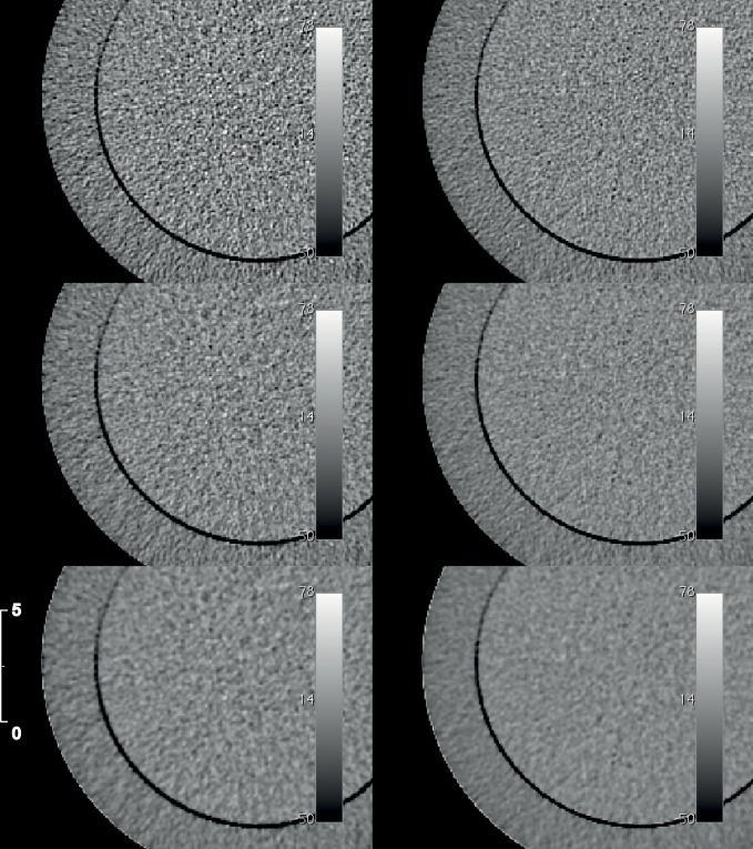
Background: Hybrid imaging combines nuclear medicine imaging such as single photon emission computed tomography (SPECT) or positron emission tomography (PET) with computed tomography (CT). Through this hybrid design, scanned patients accumulate radiation exposure from both applications. Imaging modalities have been the subject of long-term optimization efforts, focusing on diagnostic applications. It was the aim of this study to investigate the influence of an iterative CT image reconstruction algorithm (ASIR) on the image quality of the low-dose CT images.
Methodology/principal findings: Examinations were performed with a SPECT-CT scanner with standardized CT and SPECT-phantom geometries and CT protocols with systematically reduced X-ray tube currents. Analyses included image quality with respect to photon flux. Results were compared to the standard FBP reconstructed images. The general impact of the CT-based attenuation maps used during SPECT reconstruction was examined for two SPECT phantoms. Using ASIR for image reconstructions, image noise was reduced compared to FBP reconstructions for the same X-ray tube current. The Hounsfield unit (HU) values reconstructed by ASIR were correlated to the FBP HU values(R2 ≥ 0.88) and the contrast-to-noise ratio (CNR) was improved by ASIR. However, for a phantom with increased attenuation, the HU values shifted for low X-ray tube currents I ≤ 60 mA (p ≤ 0.04). In addition, the shift of the HU values was observed within the attenuation corrected SPECT images for very low X-ray tube currents (I ≤ 20 mA, p ≤ 0.001).
Conclusion/significance: In general, the decrease in X-ray tube current up to 30 mA in combination with ASIR led to a reduction of CT-related radiation exposure without a significant decrease in image quality.
Conflict of interest statement
Figures


Similar articles
-
Initial phantom study comparing image quality in computed tomography using adaptive statistical iterative reconstruction and new adaptive statistical iterative reconstruction v.J Comput Assist Tomogr. 2015 May-Jun;39(3):443-8. doi: 10.1097/RCT.0000000000000216. J Comput Assist Tomogr. 2015. PMID: 25654782
-
Correction of photon attenuation and collimator response for a body-contouring SPECT/CT imaging system.J Nucl Med. 2005 May;46(5):868-77. J Nucl Med. 2005. PMID: 15872362 Clinical Trial.
-
A comparative study based on image quality and clinical task performance for CT reconstruction algorithms in radiotherapy.J Appl Clin Med Phys. 2016 Jul 8;17(4):377-390. doi: 10.1120/jacmp.v17i4.5763. J Appl Clin Med Phys. 2016. PMID: 27455472 Free PMC article.
-
[Incident Photon Number and Reconstructed Linear Attenuation Coefficients in Iterative CT Image Reconstruction].Igaku Butsuri. 2019;38(4):143-158. doi: 10.11323/jjmp.38.4_143. Igaku Butsuri. 2019. PMID: 30828046 Review. Japanese.
-
Improvement of image resolution and quantitative accuracy in clinical Single Photon Emission Computed Tomography.Comput Med Imaging Graph. 2001 Mar-Apr;25(2):135-46. doi: 10.1016/s0895-6111(00)00064-1. Comput Med Imaging Graph. 2001. PMID: 11137790 Review.
Cited by
-
Improvement of image quality and dose management in CT fluoroscopy by iterative 3D image reconstruction.Eur Radiol. 2017 Sep;27(9):3625-3634. doi: 10.1007/s00330-017-4754-7. Epub 2017 Feb 6. Eur Radiol. 2017. PMID: 28168371
-
Radiology's Ionising Radiation Paradox: Weighing the Indispensable Against the Detrimental in Medical Imaging.Cureus. 2023 Jul 10;15(7):e41623. doi: 10.7759/cureus.41623. eCollection 2023 Jul. Cureus. 2023. PMID: 37435015 Free PMC article. Review.
-
The role of activity, scan duration and patient's body mass index in the optimization of FDG imaging protocols on a TOF-PET/CT scanner.EJNMMI Phys. 2021 Apr 6;8(1):35. doi: 10.1186/s40658-021-00380-9. EJNMMI Phys. 2021. PMID: 33825058 Free PMC article.
-
Reducing the number of CTs performed to monitor personalized dosimetry during peptide receptor radionuclide therapy (PRRT).EJNMMI Phys. 2018 Jun 19;5(1):10. doi: 10.1186/s40658-018-0211-1. EJNMMI Phys. 2018. PMID: 29916115 Free PMC article.
-
Optimization of SPECT/CT imaging protocols for quantitative and qualitative 99mTc SPECT.EJNMMI Phys. 2021 Jul 30;8(1):57. doi: 10.1186/s40658-021-00405-3. EJNMMI Phys. 2021. PMID: 34328565 Free PMC article.
References
-
- Weber WA (2005) Use of PET for monitoring cancer therapy and for predicting outcome. Journal of Nuclear Medicine 46: 983–995. - PubMed
Publication types
MeSH terms
LinkOut - more resources
Full Text Sources
Other Literature Sources
Medical
Miscellaneous
