. 2015 Sep 21;10(9):e0138607.
doi: 10.1371/journal.pone.0138607. eCollection 2015.

Aberrant Apoptotic Response of Colorectal Cancer Cells to Novel Nucleoside Analogues

Affiliations

Aberrant Apoptotic Response of Colorectal Cancer Cells to Novel Nucleoside Analogues

Leonie Harmse et al. PLoS One. .

Abstract

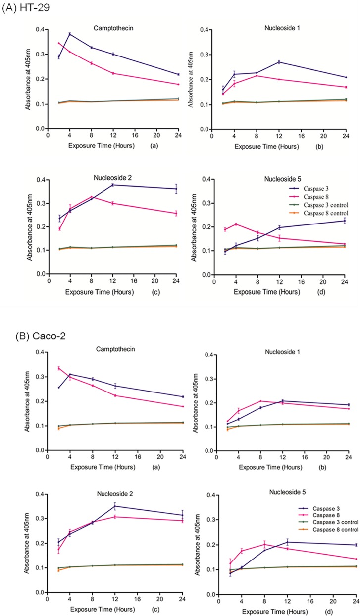
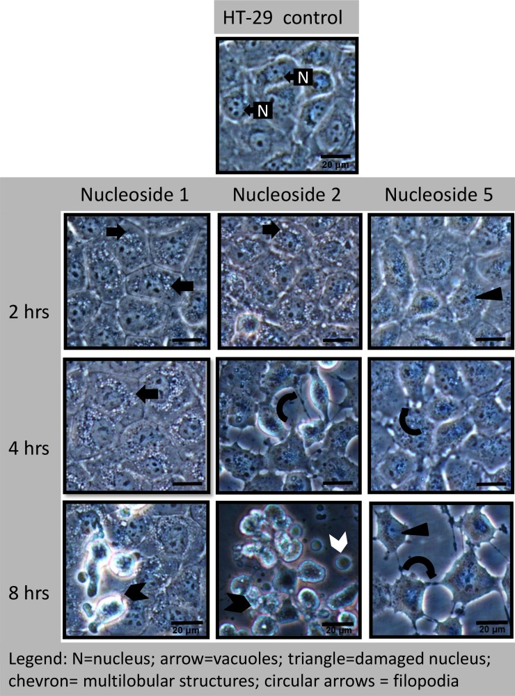
Despite the increased understanding of colorectal cancer and the introduction of targeted drug therapy, the metastatic phase of the disease remains refractory to treatment. Since the deregulation of normal apoptosis contributes to the pathogenesis of colorectal cancer, novel nucleoside analogues were synthesized here and evaluated for their ability to induce apoptosis and cause cell death in two colorectal adeno-carcinoma cell lines, Caco-2 and HT-29. Three novel nucleoside analogues assessed here showed cytotoxic activity, as measured by the MTT assay against both cell lines: the IC50 values ranged between 3 and 37 μM, with Caco-2 cells being more sensitive than HT-29 cells. Compared to camptothecin, the positive control, the nucleoside analogues were significantly less toxic to normal unstimulated leukocytes (p>0.05). Moreover, the nucleosides were able to induce apoptosis as measured by an increase in caspase 8 and caspase 3 activity above that of the control. This was additionally supported by data derived from Annexin V-FITC assays. Despite marginal changes to the mitochondrial membrane potential, all three nucleosides caused a significant increase in cytosolic cytochrome c (p>0.05), with a corresponding decrease in mitochondrial cytochrome c. Morphological analysis of both cell lines showed the rapid appearance of vacuoles following exposure to two of the nucleosides, while a third caused cellular detachment, delayed cytoplasmic vacuolisation and nuclear abnormalities. Preliminary investigations, using the autophagic indicator monodansylcadaverine and chloroquine as positive control, showed that two of the nucleosides induced the formation of autophagic vacuoles. In summary, the novel nucleoside analogues showed selective cytotoxicity towards both cancer cell lines and are effective initiators of an unusual apoptotic response, demonstrating their potential to serve as structural scaffolds for more potent analogues.


Conflict of interest statement

Competing Interests: The authors have declared that no competing interests exist.

Figures

Fig 1
Fig 1. Structures of the active nucleoside analogues (A), their respective IC50 values compared to camptothecin, in the Caco-2 and HT-29 cell lines (B), and (C) the leucocyte survival bargraph. (B) The bars represent the mean ± SEM of the IC50 values from four MTT assays carried out for each test nucleoside. The HT-29 and Caco-2 cells were exposed to the nucleoside analogues at concentrations ranging from 1 μM to 120 μM. Normal peripheral leukocytes (C) were exposed to 100 μM of nucleoside 1, 2, 5 and camptothecin for 24 hrs.
Fig 2
Fig 2. Active nucleosides induce caspase 3 and caspase 8 activity in both HT-29 (A) and in Caco-2 (B) cells after various exposure periods to 100 μM camptothecin (a), nucleoside 1 (b), nucleoside 2 (c), and nucleoside 5 (d) Data points on the graphs represent the mean of the triplicate values of caspase activity after cells were exposed to the nucleoside analogues for the indicated time periods.
Fig 3
Fig 3. Nucleosides 1, 2 and 5 cause the redistribution of cytochrome c from mitochondria to the cytosol in HT-29 (A) and Caco-2 (B).
The cells were exposed to 100 μM of nucleoside 1, 2, 5 and camptothecin, respectively, for 24 hours. The bars represent the mean ± SEM from three Elisa assays carried out for each nucleoside.
Fig 4
Fig 4. In HT-29 cells, nucleoside 1 and 2 induce nuclear fragmentation, while nucleoside 5 causes a loss of normal nuclear structure and cytoplasmic vacuolisation.
Cells were exposed to 50 μM of nucleoside 1, 2 and 5 for 24 hours and stained with Hoechst 33342 stain. Scalebar: 20 μm.
Fig 5
Fig 5. In Caco-2 cells, nucleoside 1 and 2 cause perinuclear vacuole formation, while nucleoside 5 causes a loss of normal nuclear structure, nuclear budding and cytoplasmic vacuolisation.
Cells were exposed to 50 μM of nucleoside 1, 2 and 5 for 24 hours and stained with Hoechst 33342 stain. Scalebar: 20 μm.
Fig 6
Fig 6. Nucleoside-induced morphological changes in HT-29 cells include rapid perinuclear vacuole formation (nucleoside 1 and 2) and a loss of cellular adherence (nucleoside 5).
Cells were exposed to 50 μM of the nucleoside for the indicated time periods. Experiments were repeated three times. Scale bar: 20 μm.
Fig 7
Fig 7. Nucleoside-induced morphological changes in Caco-2 cells include rapid perinuclear vacuole formation (nucleoside 1 and 2) and a loss of cellular adherence (nucleoside 5).
Cells were exposed to 50 μM of each nucleoside for the indicated times and the experiments were repeated three times. Scalebar: 20 μm.
Fig 8
Fig 8. Nucleoside 2 treated vacuolated HT-29 and Caco-2 cells stained with acridine orange shows an absence of lysosomal staining (A) and a nucleoside 2 induced HT-29 multi-lobular structure (B).
Acridine orange stained HT-29 and Caco-2 cells, following exposure to 50 μM of nucleoside 2 (A). HT-29 cells were exposed to 50 μM of nucleoside 1 and the supernatant harvested and centrifuged at 500 x g for 5 mins. The multi-lobular cell was stained with Hoechst 33342 and acridine orange (B). Scalebar: 20 μm.
Fig 9
Fig 9. Nucleoside 5 causes aggregation of actin in HT-29 cells, but not in Caco-2 cells.
Cells were treated with 50 μM of nucleoside 5 for 6 hours. Scalebar: 20μm.
Fig 10
Fig 10. Vacuoles observed under phase contrast microscopy do not co-localize with MDC stained fluorescent granules.
HT-29 cells were exposed to 50 μM of nucleoside 2 for 3 hours before MDC staining.
Fig 11
Fig 11. Nucleoside 2 and chloroquine cause the formation of large, highly fluorescent autophagic vacuoles in Caco-2 cells.
Cells were exposed to nucleoside 1, 2 and 5 at a concentration of 50 μM for three hours. Chloroquine (50 μM) was used as a positive control and camptothecin (20 μM) was included as a vacuole negative control. Arrows indicate autophagic vacuoles. Scalebars: 20 μm.
Fig 12
Fig 12. Nucleoside 2 and chloroquine cause the formation of large highly fluorescent autophagic vacuoles in HT-29 cells.
Cells were exposed to nucleoside 1, 2 and 5 at a concentration of 50 μM for three hours. Chloroquine (50 μM) was used as a positive control at and camptothecin (20 μM) was included as a negative control. Arrows indicate autophagic vacuoles. Scalebars: 20 μm.

References

    1. Carrato A, Swieboda-Sadlej A, Staszewska-Skurczynska M, Lim R, Roman L, Shparyk Y, et al. Fluorouracil, leucovorin, and irinotecan plus either sunitinib or placebo in metastatic colorectal cancer: a randomized, phase III trial. Journal of Clinical Oncology. 2013; 31:1341–1347. - PubMed
    1. Huerta S, Goulet EJ, Livingston EH. Colon cancer and apoptosis. The American Journal of Surgery 2006; 191:517–526. - PubMed
    1. Yang SY, Sales KM, Fuller B, Seifalian AM, Winslet MC. Apoptosis and colorectal cancer: implications for therapy. Trends in molecular medicine 2009; 15(5):225–233. - PubMed
    1. Wong RSY. Apoptosis in cancer: from pathogenesis to treatment. Journal of Experimental & Clinical Cancer Research 2011; 30:87. - PMC - PubMed
    1. Watson A. Apoptosis and colorectal cancer. Gut 2004; 53:1701–1709. - PMC - PubMed

Publication types