Review
. 2015 Dec;72(24):4721-57.
doi: 10.1007/s00018-015-2034-8. Epub 2015 Sep 21.

Autophagy signal transduction by ATG proteins: from hierarchies to networks

Affiliations
Review

Autophagy signal transduction by ATG proteins: from hierarchies to networks

Sebastian Wesselborg et al. Cell Mol Life Sci. 2015 Dec.

Abstract

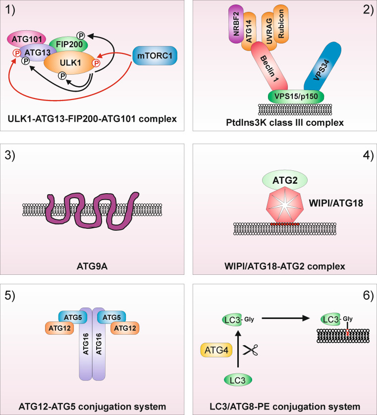
Autophagy represents an intracellular degradation process which is involved in both cellular homeostasis and disease settings. In the last two decades, the molecular machinery governing this process has been characterized in detail. To date, several key factors regulating this intracellular degradation process have been identified. The so-called autophagy-related (ATG) genes and proteins are central to this process. However, several additional molecules contribute to the outcome of an autophagic response. Several review articles describing the molecular process of autophagy have been published in the recent past. In this review article we would like to add the most recent findings to this knowledge, and to give an overview of the network character of the autophagy signaling machinery.

Keywords: ATG; Autophagy; LC3; PtdIns3K; ULK.


Conflict of interest statement

The authors declare that they have no conflict of interest.

Figures

Fig. 1
Fig. 1
Functional clusters of autophagy signaling. 1 The ULK1–ATG13–FIP200–ATG101 protein kinase complex, 2 the PtdIns3K class III complex containing the core proteins VPS34, VPS15 and Beclin 1, 3 the multi-spanning transmembrane protein ATG9A, 4 the PtdIns3P-binding WIPI/ATG18–ATG2 complex, 5 the ubiquitin-like ATG5/ATG12 system and 6 the ubiquitin-like ATG8/LC3 conjugation system. For the ULK1 complex, mTOR-dependent inhibitory phosphorylations are depicted as red arrows, and ULK1-dependent activatory phosphorylations are depicted as black arrows. For the PtdIns3K class III complex, the mutually exclusive interactions of ATG14 or UVRAG with Beclin 1 are—for simplicity—shown within one complex
Fig. 2
Fig. 2
Signaling machinery upstream and downstream of the ULK1 complex. In recent years, the mTORC1-dependent regulation of the ULK1–ATG13–FIP200–ATG101 complex has been deciphered. Under nutrient-rich conditions, mTORC1 associates with the ULK1–ATG13–FIP200–ATG101 complex and phosphorylates ULK1 and ATG13. Under starvation conditions or upon treatment with mTOR inhibitors, mTORC1 dissociates from this mega-complex, and the inhibitory mTOR-dependent phospho-sites within ULK1 and ATG13 become dephosphorylated. Active ULK1 then autophosphorylates and phosphorylates ATG13 and FIP200, ultimately leading to the initiation of autophagosome formation [, , –110]. The depicted substrates of ULK1 are listed in Table 1. MTOR has been established as central “gatekeeper” of autophagy, since this kinase integrates (1) nutrient signals, e.g., generated by growth factors or amino acids; (2) energy signals, e.g., controlled by the cellular AMP/ATP ratio; and (3) stress signals such as hypoxia or DNA damage. The Ser/Thr kinase mTOR is the catalytic subunit of two distinct kinase complexes, i.e., mTORC1 and mTORC2. The two complexes contain unique associated proteins which serve as scaffolds and determine the substrate specificity of the complexes, i.e., regulatory‐associated protein of mTOR (RAPTOR) and rapamycin‐insensitive companion of mTOR (RICTOR), respectively [–398]. Next to these two proteins, the two complexes both harbor additional specific interacting proteins and share some components. Amino acids are sensed by the RAG family of small GTPases. Active RAG heterodimers translocate mTORC1 to lysosomal surfaces, where they bind to the so-called Ragulator complex [399]. On the surface of lysosomes, mTORC1 is activated by another small GTPase termed RAS-homologue enriched in brain (RHEB). The presence of growth factors is transmitted to mTOR via AKT. AKT phosphorylates tuberous sclerosis 2 protein (TSC2; also termed tuberin), which together with TSC1 (also termed hamartin) forms the TSC1–TSC2 complex. AKT-dependent phosphorylation of TSC2 inhibits the GTPase activating protein (GAP) activity of the TSC1–TSC2 complex for RHEB, thus promoting mTORC1 activation by GTP-loaded RHEB [–402]. Alternatively, AKT phosphorylates PRAS40, which is subsequently bound by 14-3-3 proteins and cannot inhibit mTORC1 anymore [–405]. Low energy levels as sensed by a high AMP/ATP ratio are transmitted to mTORC1 via AMPK. AMPK can—like AKT—phosphorylate TSC2. However, AMPK-dependent TSC2 phosphorylation leads to increased GAP activity of the TSC1-TSC2 complex and thus to mTORC1 inhibition [406, 407]. Alternatively, AMPK can directly inhibit mTORC1 by RAPTOR phosphorylation [137]. Stress signals like hypoxia, DNA damage, TRAIL or Ca2+ signals also inhibit mTORC1 via AMPK and/or the TSC1‐TSC2 complex (reviewed in [42, 126, 127]). Finally, AKT and AMPK can directly regulate ULK1, and ULK1 can—by negative feedback loops—regulate the upstream kinases mTORC1 and AMPK [120, 121, 128, 140, 141, 143, 153, 154]
Fig. 3
Fig. 3
Signaling machinery upstream and downstream of the PtdIns3K class III complex. The PtdIns3K class III core complex consists of the catalytic subunit VPS34, the adaptor VPS15 (p150), and Beclin 1 (ATG6). Beclin 1 binds to additional regulatory proteins, including ATG14, NRBF2, UBRAG, Rubicon, AMBRA1, BCL2, and several others (reviewed in [–175]). Furthermore, there exists considerable crosstalk between the ULK1-complex and the PtdIns3K class III complex (for details see “The PtdIns3K class III complex”). ULK1 phosphorylates Beclin 1, AMBRA1, and VPS34. In turn, AMBRA1 regulates ULK1 stability and activation. Next to ULK1 itself, several ULK1-regulating kinases—such as mTORC1, AMPK, and AKT—also regulate the PtdIns3K class III complex. The product of the PtdIns3K class III catalytic activity is phosphatidylinositol 3-phosphate (PtdIns3P) (red circles). PtdIns3P then recruits the downstream effectors DFCP1 and proteins of the WIPI-family. For simplicity, the mutually exclusive interactions of ATG14 or UVRAG with Beclin 1 are shown within one complex
Fig. 4
Fig. 4
The ATG “spiderweb”. This scheme depicts the crosstalk between the six ATG signaling modules described in this review. The adjacent positioning of proteins within the single modules does not necessarily reflect direct interactions of the components. Lines can indicate both interaction and/or phosphorylation (by ULK1). Crosstalks identified for yeast orthologs are indicated by red lines

References

    1. Ashford TP, Porter KR. Cytoplasmic components in hepatic cell lysosomes. J Cell Biol. 1962;12:198–202. doi: 10.1083/jcb.12.1.198. - DOI - PMC - PubMed
    1. Novikoff AB, Essner E. Cytolysomes and mitochondrial degeneration. J Cell Biol. 1962;15:140–146. doi: 10.1083/jcb.15.1.140. - DOI - PMC - PubMed
    1. Klionsky DJ. Autophagy revisited: a conversation with Christian de Duve. Autophagy. 2008;4(6):740–743. doi: 10.4161/auto.6398. - DOI - PubMed
    1. Choi AM, Ryter SW, Levine B. Autophagy in human health and disease. N Engl J Med. 2013;368(7):651–662. doi: 10.1056/NEJMra1205406. - DOI - PubMed
    1. Levine B, Kroemer G. Autophagy in the pathogenesis of disease. Cell. 2008;132(1):27–42. doi: 10.1016/j.cell.2007.12.018. - DOI - PMC - PubMed

Publication types

Substances

LinkOut - more resources