. 2016;13(1):84-96.
doi: 10.2174/1567205012666150921100515.

Effects of Maternal Choline Supplementation on the Septohippocampal Cholinergic System in the Ts65Dn Mouse Model of Down Syndrome

Affiliations

Effects of Maternal Choline Supplementation on the Septohippocampal Cholinergic System in the Ts65Dn Mouse Model of Down Syndrome

Christy M Kelley et al. Curr Alzheimer Res. 2016.

Abstract

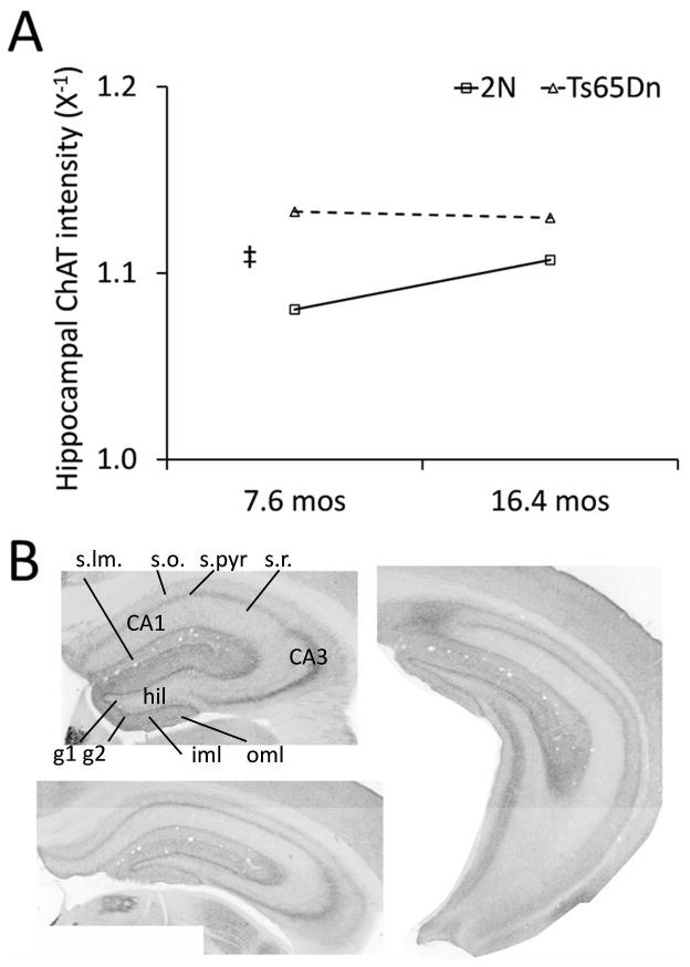
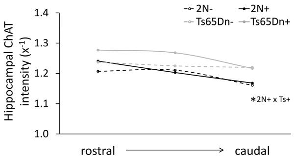
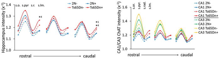
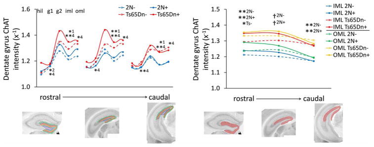
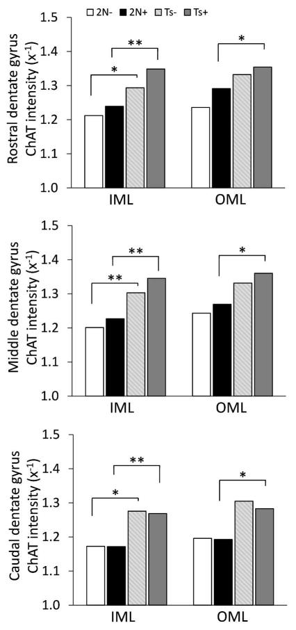
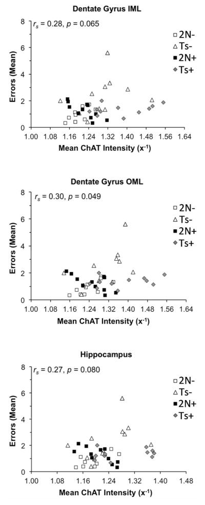
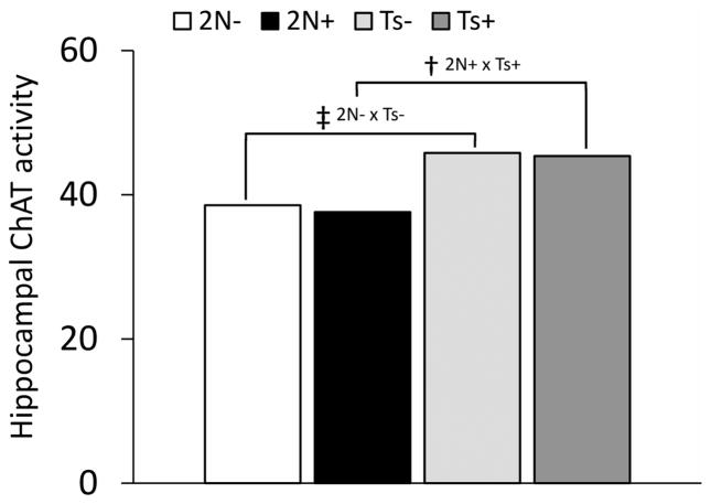
Down syndrome (DS), caused by trisomy of chromosome 21, is marked by intellectual disability (ID) and early onset of Alzheimer's disease (AD) neuropathology including hippocampal cholinergic projection system degeneration. Here we determined the effects of age and maternal choline supplementation (MCS) on hippocampal cholinergic deficits in Ts65Dn mice compared to 2N mice sacrificed at 6-8 and 14-18 months of age. Ts65Dn mice and disomic (2N) littermates sacrificed at ages 6-8 and 14-18 mos were used for an aging study and Ts65Dn and 2N mice derived from Ts65Dn dams were maintained on either a choline-supplemented or a choline-controlled diet (conception to weaning) and examined at 14-18 mos for MCS studies. In the latter, mice were behaviorally tested on the radial arm Morris water maze (RAWM) and hippocampal tissue was examined for intensity of choline acetyltransferase (ChAT) immunoreactivity. Hippocampal ChAT activity was evaluated in a separate cohort. ChAT-positive fiber innervation was significantly higher in the hippocampus and dentate gyrus in Ts65Dn mice compared with 2N mice, independent of age or maternal diet. Similarly, hippocampal ChAT activity was significantly elevated in Ts65Dn mice compared to 2N mice, independent of maternal diet. A significant increase with age was seen in hippocampal cholinergic innervation of 2N mice, but not Ts65Dn mice. Degree of ChAT intensity correlated negatively with spatial memory ability in unsupplemented 2N and Ts65Dn mice, but positively in MCS 2N mice. The increased innervation produced by MCS appears to improve hippocampal function, making this a therapy that may be exploited for future translational approaches in human DS.


Conflict of interest statement

CONFLICT OF INTEREST

The authors have no conflicts of interest to disclose.

Figures

Figure 1
Figure 1. Average ChAT intensity in the hippocampus and dentate gyrus of male Ts65Dn and 2N mice at 6–8 and 14–18 mos
(A) Ts65Dn mice had overall higher levels of cholinergic innervation than 2N mice, as measured by ChAT-immunolabeling intensity, and 2N mice showed a significant increase with age (* p < 0.05, ‡ p < 0.001, Mann-Whitney U, two-tail, n = 10–13) Values are plotted as reciprocal (x−1) of luminosity and were derived by averaging measurements in all laminae and across the three sections along the rostrocaudal access used for evaluation, shown in B. Data presented represent medians for group.
Figure 2
Figure 2. Average ChAT-positive fiber innervation in the hippocampus of MCS (+) and unsupplemented (−) male Ts65Dn and 2N mice at 14–18 mos
ChAT intensity averaged across all regions and laminae is shown as medians for each group and location. Ts65Dn supplemented mice had consistently higher values of ChAT intensity than 2N supplemented mice, significant in the caudal section (* p < 0.05, Mann-Whitney U, two-tail, n = 11 for each group). Values are plotted as reciprocal (x−1) of luminosity measures.
Figure 3
Figure 3. Cholinergic innervation to the hippocampus proper laminae in MCS (+) and unsupplemented (−) male Ts65Dn mice and disomic littermates (2N) at 14–18 mos
Line plots traversing the rostral through caudal hippocampus with measurements taken at laminae of the CA region were drawn using group medians for measurements at the stratum oriens (s.o.), stratum pyramidale (s.pyr), stratum radiatum (s.r.), and stratum lacunosum moleculare (s.lm.). (Left panel) Similar patterns of innervation were observed between groups in all hippocampal laminae (* p < 0.05, ** p < 0.01; 1, 2N-compared with Ts65Dn−; 4, 2N+ compared with Ts65Dn+, Mann-Whitney U, two-tail, n = 11 per group). (Right panel) ChAT intensity was consistently higher in CA3 than CA1, (significance p < 0.05 or greater for all groups, not shown, Friedman test). All values plotted are reciprocal (x−1) values of luminosity measurements with light-microscope photomicrographs. CA1, cornu ammonis 1; CA3, cornu ammonis 3.
Figure 4
Figure 4. Cholinergic innervation to the dentate gyrus in MCS (+) and unsupplemented (−) male Ts65Dn mice and disomic littermates (2N) at 14–18 mos
Line plots depict median ChAT luminosity measurements from the rostral hippocampus through the caudal hippocampus, with measurements taken at the hilus (hil), granule layer low density (g1), granule layer high density (g2), and dentate gyrus inner molecular layer (iml) and outer molecular layer (oml). (Left panel) Laminae-dependent alterations to the dentate gyrus were observed with maternal choline supplementation (* p < 0.05, ** p < 0.01; 1, 2N- compared with Ts65Dn−; 2, 2N- compared with 2N+, 4, 2N+ compared with Ts65Dn+, Mann-Whitney U, two-tail, n = 11 per group), and (Right panel) comparison of the inner molecular layer (IML) with the outer molecular layer (OML) shows consistently higher innervation to the OML for all groups (* p < 0.05, ** p < 0.01, † p < 0.001, Friedman test, two-tail, n = 11 per group). All values plotted represent group medians and are reciprocal (x−1) values of luminosity measurements with light-microscope photomicrographs.
Figure 5
Figure 5. Cholinergic innervation to the molecular layers of the dentate gyrus in MCS (+) and unsupplemented (−) male Ts65Dn mice and disomic littermates (2N) at 14–18 mos
Elevated ChAT intensity is seen in the inner molecular layer (IML) of Ts65Dn mice compared with 2N mice independent of treatment condition, but only in the outer molecular layer (OML) for groups that received MCS (* p < 0.05, ** p < 0.01; Mann-Whitney U, two-tail, n = 11 per group). All values represent medians and are plotted as reciprocal (x−1) values of luminosity measurements derived using light-microscope photomicrographs.
Figure 6
Figure 6. Correlations between behavioral performance on the RAWM and ChAT intensity levels in the hippocampus
Mean of errors in representative block 3 of the hidden platform task positively correlated with the intensity of ChAT immunolabeling in the inner molecular layer (IML) and outer molecular layer (OML) of the dentate gyrus as well as in the hippocampus proper for maternal choline supplemented (+) and unsupplemented (−) male Ts65Dn (Ts) mice and disomic (2N) littermates at 14–18 mos. rs, Spearman’s rank correlation coefficient
Figure 7
Figure 7. Hippocampal ChAT enzyme activity in MCS (+) and unsupplemented (−) male Ts65Dn mice and disomic littermates (2N) at 15–19 mos
ChAT activity is elevated in the hippocampus of Ts65Dn mice compared with age-matched 2N mice, independent of maternal diet treatment group († p < 0.005, ‡ p < 0.001; Mann-Whitney U, two-tail, n = 15–18 per group). Values plotted are representative of group medians.

References

    1. Määttä T, Tervo-Määttä T, Taanila A, Kaski M, Iivanainen M. Mental health, behaviour and intellectual abilities of people with Down syndrome. Downs Syndr Res Pract. 2006;11(1):37–43. - PubMed
    1. Epstein CJ. Epilogue: toward the twenty-first century with Down syndrome--a personal view of how far we have come and of how far we can reasonably expect to go. Prog Clin Biol Res. 1995;393:241–6. - PubMed
    1. Mufson EJ, Counts SE, Perez SE, Ginsberg SD. Cholinergic system during the progression of Alzheimer’s disease: therapeutic implications. Expert Rev Neurother. 2008;8(11):1703–18. - PMC - PubMed
    1. Sendera TJ, Ma SY, Jaffar S, Kozlowski PB, Kordower JH, Mawal Y, et al. Reduction in TrkA-immunoreactive neurons is not associated with an overexpression of galaninergic fibers within the nucleus basalis in Down’s syndrome. J Neurochem. 2000;74(3):1185–96. - PubMed
    1. Mesulam MM, Mufson EJ, Wainer BH, Levey AI. Central cholinergic pathways in the rat: an overview based on an alternative nomenclature (Ch1–Ch6) Neuroscience. 1983;10(4):1185–201. - PubMed

Publication types

MeSH terms