. 2015 Sep 22:6:8379.
doi: 10.1038/ncomms9379.

Circularly polarized light detection with hot electrons in chiral plasmonic metamaterials

Affiliations

Circularly polarized light detection with hot electrons in chiral plasmonic metamaterials

Wei Li et al. Nat Commun. .

Abstract

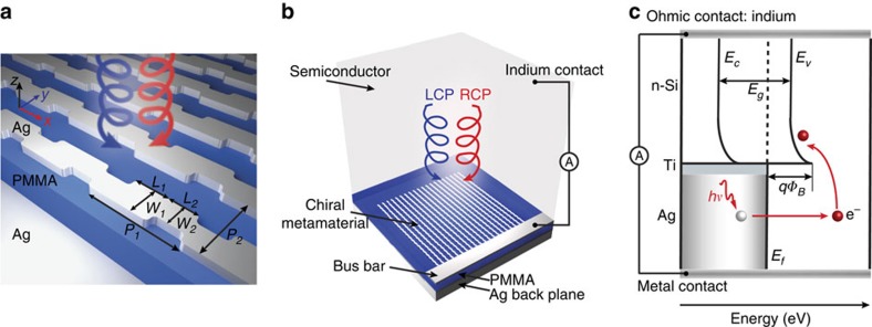
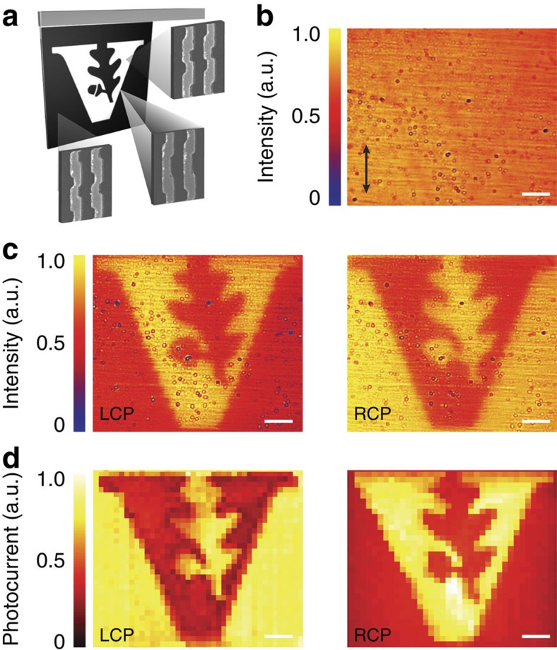
Circularly polarized light is utilized in various optical techniques and devices. However, using conventional optical systems to generate, analyse and detect circularly polarized light involves multiple optical elements, making it challenging to realize miniature and integrated devices. While a number of ultracompact optical elements for manipulating circularly polarized light have recently been demonstrated, the development of an efficient and highly selective circularly polarized light photodetector remains challenging. Here we report on an ultracompact circularly polarized light detector that combines large engineered chirality, realized using chiral plasmonic metamaterials, with hot electron injection. We demonstrate the detector's ability to distinguish between left and right hand circularly polarized light without the use of additional optical elements. Implementation of this photodetector could lead to enhanced security in fibre and free-space communication, as well as emission, imaging and sensing applications for circularly polarized light using a highly integrated photonic platform.


Figures

Figure 1
Figure 1. Schematic of the chiral metamaterial and the CPL detector.
(a) Schematic of the chiral metamaterial consisting of the chiral plasmonic meta-molecule array, dielectric spacer and metal backplane. The dimensions of the meta-molecules are L1=125 nm, L2=105 nm, W1=115 nm, W2=85 nm, P1=335 nm and P2=235 nm. The thickness of the meta-molecules, dielectric spacer and the metal backplane are 40, 160 and 100 nm, respectively. (b) Schematic of the CPL detector consisting of a chiral metamaterial integrated with a semiconductor that serves as a hot electron acceptor. The Ohmic contact on Si is formed by soldering indium. The circuit is formed by wire bonding to the silver bus bar and indium. (c) Energy band diagram of the CPL detector. A Schottky barrier is formed between Si and the Ti interfacial layer. The hot electrons that are photo-generated in the Ag metamaterial are then injected over this barrier into the Si.
Figure 2
Figure 2. Simulated optical response of chiral metamaterial.
(a,b) Simulated optical absorption spectra under LCP (blue) and RCP (red) illumination for LH (a) and RH (b) metamaterials. (c) Circular dichroism spectra for both the LH (blue) and RH (red) metamaterial. (d) CD as a function of resonator size. Dimensions of the structures (I–V) are follows: L1=115, 125, 130, 150 and 160 nm; L2=95, 105, 120, 130 and 140 nm; W1=110, 115, 120, 120 and 140 nm; W2=85, 85, 90, 90 and 100 nm; P1=305, 335, 370, 410 and 440 nm; P2=230, 235, 240, 240 and 260 nm, respectively. The other dimensions are the same as Fig. 1a. (e,f) For the LH metamaterial, the reflected LPL components, Ex (red) and Ey (blue), on multiple reflections for LCP (e) and RCP (f) light at wavelength of 1,350 nm. (g,h) Simulated electric (g) and magnetic (h) fields for LCP and RCP illumination at wavelength of 1,350 nm.
Figure 3
Figure 3. Experimentally measured optical absorption and photoresponsivity spectra.
(a,b) Scanning electron microscope images of the LH (a) and RH (b) metamaterial before spin coating the PMMA spacer layer. The inset shows a unit cell of the chiral metamaterial. Scale bar, 500 nm. (c) Schematic of experimental set-up. (d,e) Experimentally measured optical absorption spectra under LCP (blue) and RCP (red) illumination for LH (d) and RH (e) metamaterials. (f) Experimentally measured circular dichroism spectra for both LH (blue) and RH (red) metamaterials. (g,h) Experimentally measured (dots) and theoretically calculated (solid curve) photoresponsivity spectra under LCP (blue) and RCP (red) illumination for LH (g) and RH (h) metamaterials. (i) Photocurrent polarization discrimination ratio spectra of LH and RH metamaterials. The metamaterials measured have overall areas of 70 × 70 μm2.
Figure 4
Figure 4. Spatial scanning and bias-dependent photocurrent.
(a) Scanning photocurrent map of LH (top) and RH (bottom) metamaterials under LCP and RCP illumination. Scale bar, 15 μm. (b) Bias dependency of photocurrent of a LH metamaterial for LCP (blue) and RCP (red) light under a laser power of 1.5 mW. The green curve shows the polarization discrimination ratio versus bias.
Figure 5
Figure 5. CPL detector with RH and LH elements patterned into the Vanderbilt University logo.
(a) Schematic of the pattern with the LH metamaterial filling the black region and the RH metamaterial filling the white region. (b) Camera image of the metamaterial under linearly polarized light with polarization along the vertical direction. (c) Camera images of the metamaterial under LCP (left) and RCP (right) illumination. (d) Scanning photocurrent maps of the metamaterial under LCP (left) and RCP (right) illumination. Scale bar, 10 μm.

References

    1. Sherson J. F. et al. Quantum teleportation between light and matter. Nature 443, 557–560 (2006). - PubMed
    1. Wagenknecht C. et al. Experimental demonstration of a heralded entanglement source. Nat. Photon. 4, 549–552 (2010).
    1. Togan E. et al. Quantum entanglement between an optical photon and a solid-state spin qubit. Nature 466, 730–734 (2010). - PubMed
    1. Farshchi R., Ramsteiner M., Herfort J., Tahraoui A. & Grahn H. T. Optical communication of spin information between light emitting diodes. Appl. Phys. Lett. 98, 162508 (2011).
    1. Greenfield N. J. Using circular dichroism spectra to estimate protein secondary structure. Nat. Protoc. 1, 2876–2890 (2006). - PMC - PubMed

Publication types