. 2015 Sep 22:6:8277.
doi: 10.1038/ncomms9277.

Atomic description of the immune complex involved in heparin-induced thrombocytopenia

Affiliations

Atomic description of the immune complex involved in heparin-induced thrombocytopenia

Zheng Cai et al. Nat Commun. .

Abstract

Heparin-induced thrombocytopenia (HIT) is an autoimmune thrombotic disorder caused by immune complexes containing platelet factor 4 (PF4), antibodies to PF4 and heparin or cellular glycosaminoglycans (GAGs). Here we solve the crystal structures of the: (1) PF4 tetramer/fondaparinux complex, (2) PF4 tetramer/KKO-Fab complex (a murine monoclonal HIT-like antibody) and (3) PF4 monomer/RTO-Fab complex (a non-HIT anti-PF4 monoclonal antibody). Fondaparinux binds to the 'closed' end of the PF4 tetramer and stabilizes its conformation. This interaction in turn stabilizes the epitope for KKO on the 'open' end of the tetramer. Fondaparinux and KKO thereby collaborate to 'stabilize' the ternary pathogenic immune complex. Binding of RTO to PF4 monomers prevents PF4 tetramerization and inhibits KKO and human HIT IgG-induced platelet activation and platelet aggregation in vitro, and thrombus progression in vivo. The atomic structures provide a basis to develop new diagnostics and non-anticoagulant therapeutics for HIT.


Conflict of interest statement

The authors declare no competing financial interest but a patent has been filed on the use of the antibodies for HIT by the University of Pennsylvania. Zheng Cai, Douglas B. Cines, Zhiqiang Zhu and Mark I. Greene are listed as inventors.

Figures

Figure 1
Figure 1. Crystal structure of the fondaparinux/PF4 tetramer complex.
(a) Overall structure of the PF4/fondaparinux complex. Fondaparinux makes contacts with a single PF4 tetramer in the groove among the monomers on one side of the asymmetric tetramer. Monomers A, B, C and D in one PF4 tetramer are coloured in green, cyan, magenta and yellow, respectively. (b) Fondaparinux (stick representations) stabilizes the PF4 tetramer by binding in the groove among three monomers in a PF4 tetramer. Yellow dotted lines indicate the polar interactions between fondaparinux and three PF4 monomers. (c) One fondaparinux (spheres) denoted in the blue box binds in the groove of one tetramer (cartoon representation on the left) and also binds to the C-terminal helix of a second tetramer (cartoon representation on the right), thereby bridging PF4 tetramers. (d) Electrostatic potential surface representation (positive: blue; negative: red) of the PF4 tetramer shows that fondaparinux binds along a continuous positively charged surface on the ‘closed' side of PF4 tetramer. (e) Detailed representation of the positively charged residues (coloured in blue and labelled) on the fondaparinux binding interface between two PF4 tetramers. (f) Analysis of crystal lattice reveals a molecular pathway for the formation of antigenic complexes. A fragment of heparin first binds within the groove of one PF4 tetramer (limon, left); binding of the first PF4 tetramer imparts a local linearized structure on heparin, which enhances the binding of a second tetramer (pale green, middle); progression of this process eventuates in the formation of ultralarge antigenic complexes (right).
Figure 2
Figure 2. Crystal structure of the KKO-Fab/PF4 tetramer complex.
(a,b) Overall structure of the PF4/KKO-Fab complex. a: cartoon representations of the complex; b: molecular surface representations. The heavy chain and light chain of KKO-Fab are coloured in blue and light blue, respectively. (c) Detailed binding interface of HIT antibody KKO to a PF4 tetramer. Residues within a PF4 tetramer that are <5 Å away from KKO-Fab molecule are highlighted. PF4 monomers are coloured as in Fig. 1. (d,e) Binding of KKO (d) and RTO (e) to structure-based PF4 mutants. (f) Platelet aggregation by wild-type PF4 and PF4 mutants. KKO (red) induced platelet aggregation in the presence of wild-type PF4 and heparin whereas an isotype matched non-pathogenic antibody RTO described below (green) did not. The panel also demonstrates that PF4 mutants bearing mutations along the KKO binding interface (blue, PF4-9SCV; brown, PF4-55R, Cyan, PF4-9SCV55R) were unable to mediate KKO-induced platelet aggregation. (g) Model of the KKO-Fab/PF4/heparin ternary complex. Surface representations of KKO-Fab are coloured in blue (heavy chain) and light blue (light chain) and others are coloured as in Fig. 1f. The model assumes the heparin molecule is composed of about 7 structures similar to fondaparinux depicted in the figure as a non-continuous chain. Intact UFH may further enhance the stability of the holo-complex compared with the fondaparinux fragment, thereby rendering it more antigenic and more capable of binding multiple IgG antibodies.
Figure 3
Figure 3. Crystal structure of the RTO-Fab/PF4 monomer complex.
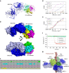
(a,b) Overall structure of the PF4/RTO-Fab complex. a: cartoon representations of the complex; b: molecular surface representations. The heavy chain and light chain of RTO-Fab are coloured in blue and light blue, respectively. (c) Detailed binding interface of the non-HIT antibody RTO to a PF4 monomer. Residues of a PF4 monomer that are <5 Å away from RTO-Fab molecule are highlighted. (d) Superposition of the PF4 monomer (green) in the RTO-Fab/PF4 complex with that in the unbound PF4 (grey) indicates that binding of RTO-Fab causes a dramatic structural change in the PF4 monomer: the C-terminal helices are shifted ∼60°. (e) Superposition of the PF4 monomer (green) in complex with RTO-Fab (blue and light blue) with the unbound PF4 tetramer (grey). The three arrows indicate the sites where binding of RTO-Fab to one PF4 monomer causes steric clashes with a second PF4 monomer in the tetramer, thereby preventing tetramer formation.
Figure 4
Figure 4. RTO prevents KKO-induced platelet activation and thrombosis.
(a). Inhibition of KKO/PF4 mediated platelet activation by RTO. Samples of whole blood were incubated with the indicated concentrations of RTO in the presence of PF4 (10 μg ml−1) for 15 min before adding the platelet-activating anti-PF4 antibody KKO or human IgG. Activation of platelets were followed by expression of P-selectin; the effect of RTO is expressed as % of geometric mean fluorescent intensity (MFI) of P-selectin expression on platelets relative to MFI in the absence of RTO. (b) In vitro platelet activation assay demonstrated that pre-incubation of PF4 with RTO prevented KKO-induced platelet aggregation. (c) Representative composite images of platelet fluorescence overlaid on brightfield snapshots of injuries in mice receiving either RTO or the IgGk2B isotype control TRA are shown. Pre KKO images show thrombi 15 min after initial injury and injection of RTO or TRA. Post KKO images represent the same thrombus 15 min after KKO had been injected intravenously. Arrows represent the direction of blood flow. (d) Each dot denotes the per cent change in the size of a single injury based on binding of fluorescently labelled platelets in mice receiving either RTO or the IgGκ2B isotype control TRA followed by KKO. Error bars show the standard deviation. N=18 injuries in three mice for RTO, N=19 injuries in three mice for TRA. P<0.0001.
Figure 5
Figure 5. Model of RTO inhibition of heparin-induced thrombocytopenia.
PF4 molecules (green circles) exist in an equilibrium among monomers, dimers and tetramers. On binding to heparin (orange circles), the configuration of the tetramer is stabilized. As a result, the open end of the PF4 tetramer is oriented and recognized by HIT-antibody KKO (black). KKO in turn fosters PF4 oligomerization and works collaboratively with heparin to stabilize the ternary complex. The net result is the generation of stable ultralarge immune complexes capable of sustained activation of Fcγ receptors on platelets and monocytes, which consequently leads to HIT. The non-HIT isotype-matched antibody RTO (blue) binds to PF4 monomers, prevents PF4 oligomerization, prevents formation of ultralarge immune complexes and as a result may prevent HIT. The cartoon assumes heparin is composed of about seven structures similar to fondaparinux.

References

    1. Linkins L. A. et al.. Treatment and prevention of heparin-induced thrombocytopenia: Antithrombotic Therapy and Prevention of Thrombosis, 9th ed: American College of Chest Physicians Evidence-Based Clinical Practice Guidelines. Chest 141, e495S–e4530 (2012). - PMC - PubMed
    1. Phillips K. W., Dobesh P. P. & Haines S. T. Considerations in using anticoagulant therapy in special patient populations. Am. J. Health Syst. Pharm. 65, S13–S21 (2008). - PubMed
    1. Lee G. M. & Arepally G. M. Heparin-induced thrombocytopenia. Hematology Am. Soc. Hematol. Educ. Program 2013, 668–674 (2013). - PMC - PubMed
    1. Lewis B. E. et al.. Argatroban anticoagulation in patients with heparin-induced thrombocytopenia. Arch. Intern. Med. 163, 1849–1856 (2003). - PubMed
    1. Cuker A. & Cines D. B. How I treat heparin-induced thrombocytopenia. Blood 119, 2209–2218 (2012). - PubMed

Publication types

MeSH terms