. 2015 Oct 19;212(11):1783-91.
doi: 10.1084/jem.20150318. Epub 2015 Sep 21.

Type 3 innate lymphoid cells maintain intestinal epithelial stem cells after tissue damage

Affiliations

Type 3 innate lymphoid cells maintain intestinal epithelial stem cells after tissue damage

Patricia Aparicio-Domingo et al. J Exp Med. .

Abstract

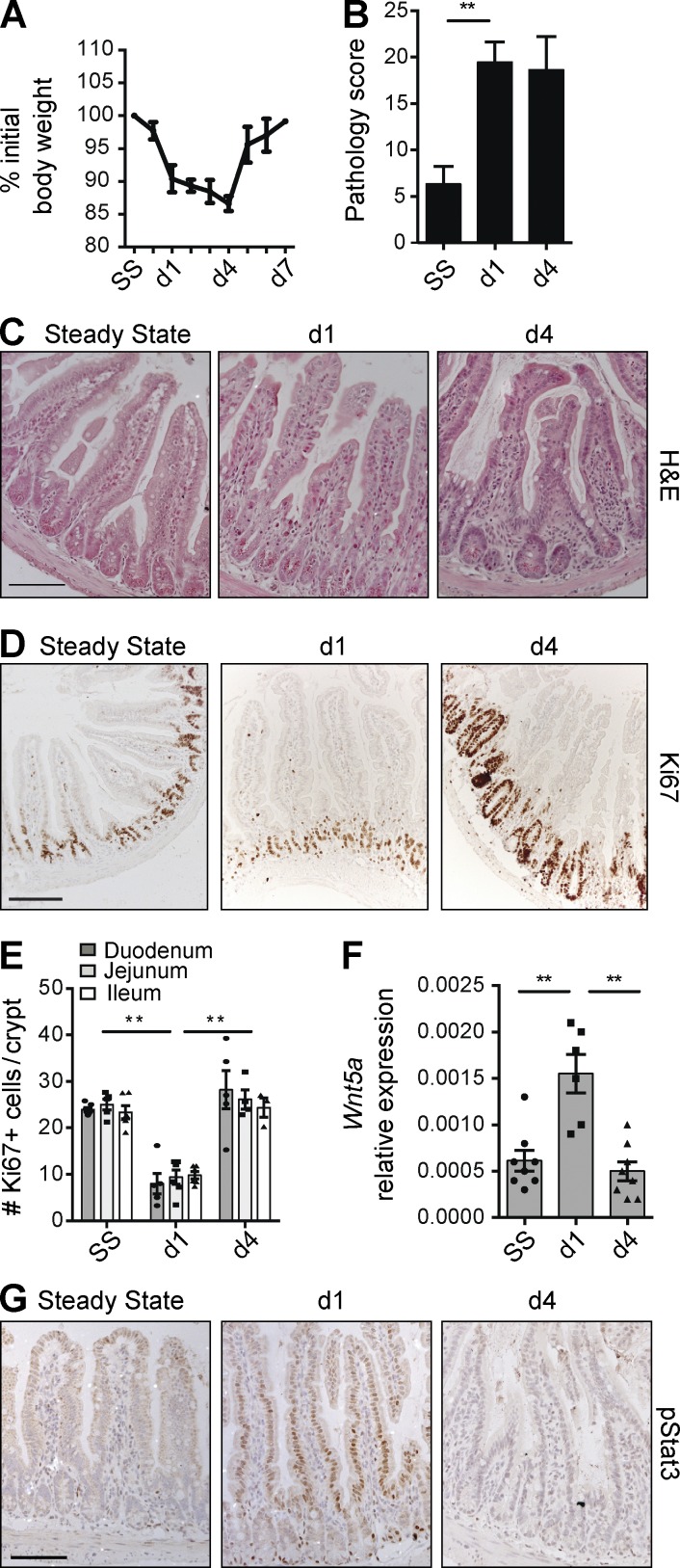
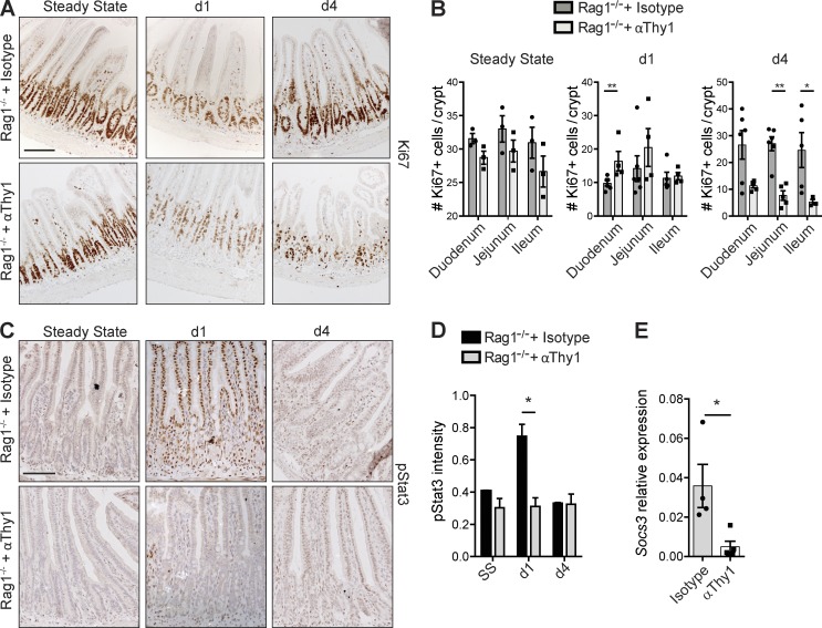
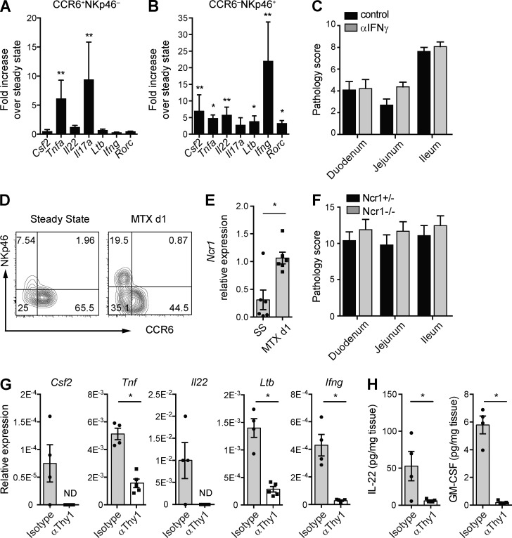
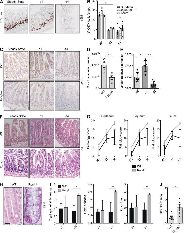
Disruption of the intestinal epithelial barrier allows bacterial translocation and predisposes to destructive inflammation. To ensure proper barrier composition, crypt-residing stem cells continuously proliferate and replenish all intestinal epithelial cells within days. As a consequence of this high mitotic activity, mucosal surfaces are frequently targeted by anticancer therapies, leading to dose-limiting side effects. The cellular mechanisms that control tissue protection and mucosal healing in response to intestinal damage remain poorly understood. Type 3 innate lymphoid cells (ILC3s) are regulators of homeostasis and tissue responses to infection at mucosal surfaces. We now demonstrate that ILC3s are required for epithelial activation and proliferation in response to small intestinal tissue damage induced by the chemotherapeutic agent methotrexate. Multiple subsets of ILC3s are activated after intestinal tissue damage, and in the absence of ILC3s, epithelial activation is lost, correlating with increased pathology and severe damage to the intestinal crypts. Using ILC3-deficient Lgr5 reporter mice, we show that maintenance of intestinal stem cells after damage is severely impaired in the absence of ILC3s or the ILC3 signature cytokine IL-22. These data unveil a novel function of ILC3s in limiting tissue damage by preserving tissue-specific stem cells.


Figures

Figure 1.
Figure 1.
Epithelial responses to MTX. (A) Weight curve of WT mice treated with MTX. (B) MTX-induced pathology as described in Materials and methods. (C and D) Representative H&E staining (C) and immunostaining of Ki67 (D) in ileal sections at the indicated time points. (E) Number of KI67+ cells per crypt at the indicated time points. (F) Transcript analysis of Wnt5a in ileal tissues. (G) Representative immunostaining of pStat3 in ileal sections at the indicated time points. (A–C and G) Two to four independent experiments, n > 5 per time point. (D–F) Two independent experiments, n = 2–3 per time point. Mean ± SEM. **, P < 0.01. Bars, 50 µm.
Figure 2.
Figure 2.
Epithelial responses to tissue damage depend on Thy1+ cells. (A and B) Representative immunostaining of Ki67 in isotype- and α-Thy1–treated Rag1−/− mice (A) and number of Ki67+ cells per crypt at the indicated time points (B). (C) Immunostaining of pStat3 in isotype- and α-Thy1–treated Rag1−/− mice. (D) pStat3 intensity in ileal sections at the indicated time points. (E) Socs3 transcript levels relative to Gapdh from ileum at day 1 after MTX (two independent experiments, n = 2–5 per time point). Mean ± SEM. *, P < 0.05; **, P < 0.01. Bars, 50 µm.
Figure 3.
Figure 3.
ILC3s are activated upon MTX-induced damage. (A and B) Fold induction of the indicated transcripts relative to steady state of lamina propria CCR6+NKp46 ILC3s (A) and CCR6NKp46+ ILC3s (B) at day 1 after MTX. (C) Pathology score at day 4 after MTX of intestines of Rag1−/− mice treated with neutralizing IFNγ or isotype control antibodies. (D) Representative flow cytometry plot of NKp46 and CCR6 expression on lamina propria ILC3s at steady state and day 1 after MTX. (E) Transcript levels of Ncr1 relative to Gapdh from sorted CCR6NKp46+ ILC3s. (F) Pathology score at day 4 after MTX of intestines of Ncr1+/− or Ncr1−/− mice. (G) Transcriptional analyses of the indicated genes relative to Gapdh from ileum of isotype control or Thy1–depleted Rag1−/− mice at day 1 after MTX. (H) Protein levels of IL-22 and GM-CSF after overnight ileal explant cultures isolated at day 1 after MTX. (A, B, D, and E) Four to six independent experiments, n = 5–7 per time point. (C and F) Two independent experiments, n = 2–4 per group. (G and H) Two independent experiments, n = 2–3 per group. Mean ± SEM. *, P < 0.05; **, P < 0.01. ND, not detected.
Figure 4.
Figure 4.
Absence of Rorγt augments crypt damage after MTX. (A) Ki67 in ileum sections of Rorγt−/− mice at the indicated time points. (B) Number of Ki67+ cells in crypts of Rorγt−/− mice at the indicated time points. (C) pStat3 in ileum sections from WT and Rorγt−/− mice at the indicated time points. (D) Socs3 transcripts relative to Gapdh from ileum of WT and Rorγt−/− mice at day 1 after MTX. (E) Wnt5a transcripts from ileum of Rorγt−/− mice at the indicated time points. (F) Representative H&E staining of ileal sections at the indicated time points. (G) MTX-induced small intestinal damage in WT and Rorγt−/− mice as specified in Materials and methods. (H) Representative high-power magnification of ileal crypts of WT and Rorγt−/− mice 4 d after MTX. (I) Crypt damage in WT and Rorγt−/− mice at days 1 and 4. (J) Ratio between bax and bcl2l1 transcript levels in ileum of WT and Rorγt−/− mice at day 1 after MTX. (A and B) Two independent experiments, n = 3–4 per group. (C, D, and F–I) Two to four independent experiments, n = 2–5 per group. (E and J) Two independent experiments, n = 2–3 per group. Mean ± SEM. *, P < 0.05; **, P < 0.01. Bars: 50 µm (A, C, and F); 10 µm (H).
Figure 5.
Figure 5.
ILC3s preserve ISCs after MTX-induced damage. (A) Percentage of GFP+ stem cells within EpCAM1+ cells from purified intestinal crypts at the indicated time points. (B) Representative H&E staining of ileal sections of WT and Rorγt−/− chimeras at day 4 after MTX. (C) Small intestinal damage in WT (black bars) and Rorγt−/− (gray bars) chimeras at day 4 after MTX as described in Materials and methods. (D) Crypt pathology score. (E) Frequency of GFP+ cells from WT and Rorγt−/− chimeras at homeostasis. (F) Representative plots of purified crypts of WT and Rorγt−/− chimeras at day 4 after MTX. Numbers adjacent to outlined areas indicate percentage of EpCAM+Lgr5-GFP+ cells. (G) Frequency of GFP+ cells from WT and Rorγt−/− chimeras at day 4 after MTX. (H) Representative plots of purified duodenal crypts at day 4 after MTX of Lgr5-GFP mice treated with aIL-22 or control antibodies. (I) Frequency of GFP+ cells at day 4 after MTX from Lgr5-GFP mice treated with aIL-22 or control antibodies. (A) Two independent experiments, n = 2 per group. (B–D, F, and G) Two independent experiments with two to four mice per group. (E) Two independent experiments, WT: n = 1 per group, KO: n = 1–3 per group. (H and I) Two independent experiments with two to five mice per group. Mean ± SEM. *, P < 0.05. Bar, 50 µm.

Comment in

References

    1. Anderson C.A., Boucher G., Lees C.W., Franke A., D’Amato M., Taylor K.D., Lee J.C., Goyette P., Imielinski M., Latiano A., et al. . 2011. Meta-analysis identifies 29 additional ulcerative colitis risk loci, increasing the number of confirmed associations to 47. Nat. Genet. 43:246–252. 10.1038/ng.764 - DOI - PMC - PubMed
    1. Artis D., and Spits H.. 2015. The biology of innate lymphoid cells. Nature. 517:293–301. 10.1038/nature14189 - DOI - PubMed
    1. Bollrath J., Phesse T.J., von Burstin V.A., Putoczki T., Bennecke M., Bateman T., Nebelsiek T., Lundgren-May T., Canli O., Schwitalla S., et al. . 2009. gp130-mediated Stat3 activation in enterocytes regulates cell survival and cell-cycle progression during colitis-associated tumorigenesis. Cancer Cell. 15:91–102. 10.1016/j.ccr.2009.01.002 - DOI - PubMed
    1. de Koning B.A., van Dieren J.M., Lindenbergh-Kortleve D.J., van der Sluis M., Matsumoto T., Yamaguchi K., Einerhand A.W., Samsom J.N., Pieters R., and Nieuwenhuis E.E.. 2006. Contributions of mucosal immune cells to methotrexate-induced mucositis. Int. Immunol. 18:941–949. 10.1093/intimm/dxl030 - DOI - PubMed
    1. Demaria M., Misale S., Giorgi C., Miano V., Camporeale A., Campisi J., Pinton P., and Poli V.. 2012. STAT3 can serve as a hit in the process of malignant transformation of primary cells. Cell Death Differ. 19:1390–1397. 10.1038/cdd.2012.20 - DOI - PMC - PubMed

Publication types