. 2015 Sep 21;16(1):201.
doi: 10.1186/s13059-015-0749-3.

Regulation of constitutive and alternative mRNA splicing across the human transcriptome by PRPF8 is determined by 5' splice site strength

Affiliations

Regulation of constitutive and alternative mRNA splicing across the human transcriptome by PRPF8 is determined by 5' splice site strength

Vihandha O Wickramasinghe et al. Genome Biol. .

Abstract

Background: Sequential assembly of the human spliceosome on RNA transcripts regulates splicing across the human transcriptome. The core spliceosome component PRPF8 is essential for spliceosome assembly through its participation in ribonucleoprotein (RNP) complexes for splice-site recognition, branch-point formation and catalysis. PRPF8 deficiency is linked to human diseases like retinitis pigmentosa or myeloid neoplasia, but its genome-wide effects on constitutive and alternative splicing remain unclear.

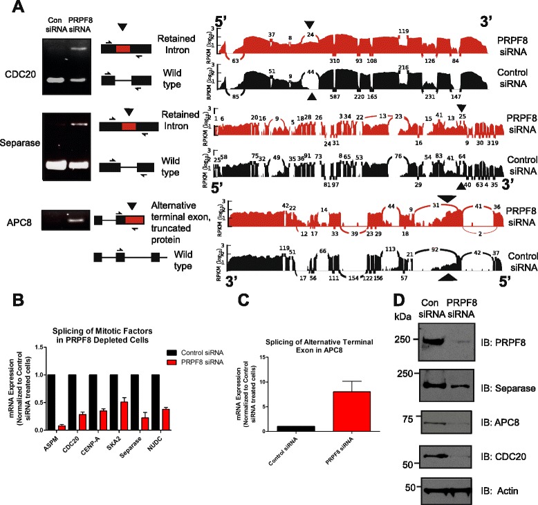
Results: Here, we show that alterations in RNA splicing patterns across the human transcriptome that occur in conditions of restricted cellular PRPF8 abundance are defined by the altered splicing of introns with weak 5' splice sites. iCLIP of spliceosome components reveals that PRPF8 depletion decreases RNP complex formation at most splice sites in exon-intron junctions throughout the genome. However, impaired splicing affects only a subset of human transcripts, enriched for mitotic cell cycle factors, leading to mitotic arrest. Preferentially retained introns and differentially used exons in the affected genes contain weak 5' splice sites, but are otherwise indistinguishable from adjacent spliced introns. Experimental enhancement of splice-site strength in mini-gene constructs overcomes the effects of PRPF8 depletion on the kinetics and fidelity of splicing during transcription.

Conclusions: Competition for PRPF8 availability alters the transcription-coupled splicing of RNAs in which weak 5' splice sites predominate, enabling diversification of human gene expression during biological processes like mitosis. Our findings exemplify the regulatory potential of changes in the core spliceosome machinery, which may be relevant to slow-onset human genetic diseases linked to PRPF8 deficiency.


Figures

Fig. 1
Fig. 1
Genome-wide decreases in spliceosome assembly at exon–intron junctions following PRPF8 depletion. a Spliceosome iCLIP reveals spliceosomal interactions with pre-mRNA that change following PRPF8 depletion at the 5′ splice site. Spliceosome iCLIP cross-link sites summarized into genome-wide maps around splice junctions reveal distinct peaks associated with individual RNA-binding protein (RBP) occupancy. A large reduction in the amplitude of the cross-link peaks located ~20 nucleotides upstream of the 5′ splice site, and immediately adjacent to the exon–intron junction following PRPF8 depletion is indicated by red arrowheads. b Spliceosome iCLIP reveals spliceosomal interactions with pre-mRNA that change following PRPF8 depletion at the 3′ splice site. A reduction in the amplitude of the cross-link peak located ~50 nucleotides upstream of the 3′ splice site following PRPF8 depletion is indicated by red arrowheads
Fig. 2
Fig. 2
Altered splicing after PRPF8 depletion affects only a subset of human transcripts. a Intronic expression levels across the genome are increased in PRPF8-depleted cells. Normalized intron expression was calculated following analysis of the transcriptome of PRPF8-depleted Cal51 cells using RNA sequencing (left; “Materials and methods”). Statistically significant pairwise comparison is indicated (***p < 0.001). The number of mapped reads, the percentage of reads that map to exons, exon–intron boundaries, and intron bodies are shown in tabular form (right). b RNA sequencing experiments demonstrate that PRPF8 depletion results in altered splicing in only a subset of human transcripts. DEXSeq (“Materials and methods”) identifies a set of 2086 protein-coding genes that contain at least one retained intron [false discovery rate (FDR) < 0.01] and a set of 1921 protein-coding genes that display significant differences in exon usage (FDR < 0.01) following PRPF8 depletion; 637 genes display both retained introns and alternative exon usage (p < 2.2e-16). Transcripts with altered splicing patterns constitute only a subset of all expressed protein-coding genes (n = 3388 out of 13,216; expression threshold = 1 FPKM (Fragments per kilobase of exon per million reads mapped); “Materials and methods”). c Functional enrichment analysis using DAVID and WebGestalt (“Materials and methods”) shows that this subset is enriched for transcripts that participate in mitosis, ubiquitin conjugation, or RNA processing. GO Gene Ontology
Fig. 3
Fig. 3
Altered splicing of transcripts encoding proteins required for mitotic progression after PRPF8 depletion. a RT-PCR analysis of RNA from PRPF8-depleted cells reveals a variety of splicing defects in transcripts that encode critical factors required for mitotic progression, including retained introns (CDC20 and Separase), and alternative terminal exons (APC8). RT-PCR analysis of CDC20, Separase, and APC8 using the primers indicated in the schematic are shown alongside the corresponding coverage plots (control siRNA in black, PRPF8 siRNA in red). The splicing defects are indicated by arrowheads. b, c Quantitative RT-PCR analysis of RNA from PRPF8-depleted cells, including retained introns (CDC20, Separase, and NUDC), skipped exons (ASPM and SKA2), and alternative terminal exons (APC8). Plots are relative to RNA levels in control siRNA-treated cells, assigned an arbitrary value of 1, and show the mean of triplicate readings from three independent experiments ± standard error of the mean. d Protein expression analysis of genes with altered splicing following PRPF8 depletion
Fig. 4
Fig. 4
PRPF8 depletion impedes mitotic progression. a PRPF8 depletion in Cal51 cells results in a fivefold increase in the percentage of mitotic cells compared with control siRNA-treated cells. Plots show the mean from three independent experiments ± standard error of the mean. Statistically significant pairwise comparison is shown (***p < 0.001). b U2OS cells stably expressing green fluorescent protein (GFP)-tagged histone H2B, depleted of PRPF8, and monitored by phase-contrast time-lapse microscopy spend >120 min in mitosis, as opposed to control siRNA-treated cells, in which mitosis lasts, on average, <60 min. The bar graph depicts time spent in mitosis, measured from nuclear envelope breakdown to anaphase. c, d Live-cell imaging of U2OS GFP-H2B cells depleted of PRPF8 reveals multiple mitotic abnormalities, including unaligned chromosomes, anaphase bridges and multipolar divisions, indicated by arrows and arrowheads. Severe defects in both chromosome alignment and segregation are also observed in some cells. Representative images are shown in (d); defects are quantified in (c). e Introduction of properly spliced and processed mRNA into PRPF8-depleted cells suffices to partially rescue the observed mitotic defects. Control siRNA-treated and PRPF8-depleted cells were transfected with properly spliced and processed mRNA and analyzed for mitotic index. Plots represent the mean from three independent experiments ± standard error of the mean. Statistically significant pairwise comparisons are shown (**p < 0.01, ***p < 0.001)
Fig. 5
Fig. 5
Depletion of Complex B components that interact directly with PRPF8 recapitulate defects in mitotic progression accompanied by the altered splicing of mitotic genes. a Depletion of Complex A components does not cause defects in either the splicing of mitotic genes (top and middle panels) or in mitotic progression (bottom panel). Independent depletion of several different components of the three major spliceosome subcomplexes (A, B, and C) was verified by qRT-PCR (Additional file 5). b Depletion of several Complex B components phenocopies PRPF8, in terms of both defects in splicing of mitotic genes (top and middle panels) and, to a lesser extent, increased mitotic index (bottom panel). Depletion of the spliceosome components BRR2 and EFTUD2, which interact directly with PRPF8 to form the U5 snRNP, most strongly phenocopy PRPF8 deficiency. c Depletion of Complex C components does not cause defects in either the splicing of mitotic genes (top and middle panels), or in mitotic progression (bottom panel). SLU7 partially phenocopies PRPF8 but does not display defects in mitotic progression. For all qRT-PCR experiments in this figure, plots are relative to RNA levels in control siRNA-treated cells, assigned an arbitrary value of 1, and show the mean of triplicate readings from at least three independent depletion experiments ± standard error of the mean (SEM). For cell cycle analysis experiments, plots represent the mean from at least three independent depletion experiments ± SEM
Fig. 6
Fig. 6
Introns that are retained and exons that are differentially used after PRPF8 depletion have weak 5′ splice sites. a Inefficiently spliced introns have weaker 5′ splice sites. A set of 200 retained introns (RI) were selected based on fold-change differences (see “Materials and methods”), and non-retained introns (NRI) within the same set of genes were used as a contrast. A 5′ splice site strength analysis was then carried out as described in “Materials and methods” for each subset. Statistically significant pairwise comparison is indicated (**p < 0.01). b Motif enrichment analysis on the same set of genes shows that although the most frequently identified motifs correspond to the consensus splice site sequences for both retained (RI) and non-retained introns (NRI), the percentage of targets with such motifs varies significantly between the two categories (68.50 % for RI versus 91.03 % for NRI; p < 2.2 × 10⁻¹⁶, Fisher’s exact test). c Analysis of GC composition reveals that retained introns have higher GC content than non-retained introns with values closer to those of adjacent exons. Differential GC content was calculated by dividing GC content of each intron to the average of its adjacent exons (see “Materials and methods”). Statistically significant pairwise comparison is indicated (***p < 0.001). d Differentially used exons have weaker 5′ splice sites. A 5′ splice site strength analysis was carried out as described in “Materials and methods” for each subset. Statistically significant pairwise comparison is indicated (***p < 0.001)
Fig. 7
Fig. 7
Enhancement of splice-site strength renders the CDC20 mini-gene resistant to PRPF8 depletion. a A mini-gene containing a full-length intron flanked by two exons within the CDC20 gene. This particular intron is retained following PRPF8 depletion as determined by RNA-sequencing experiments and the corresponding coverage plot is shown. Both the 5′ splice site and the 3′ splice site encompassing the exon–intron and intron–exon boundaries are weaker than the corresponding consensus sequences and the sequence of 5′ and 3′ splice mutant constructs are indicated with changes marked in red. RPKM Reads per kilobase of exon per million reads mapped, WT wild type. b, c Enhancement of splice site strength renders the CDC20 mini-gene resistant to PRPF8 depletion. PRPF8 depletion strongly suppresses removal of this intron in the CDC20 mini-gene (left panel). Alteration by site-directed mutagenesis of the weak 5′ splice site into a strong consensus sequence leads to efficient removal of this intron in PRPF8-depleted cells, in a manner indistinguishable from control siRNA-treated cells (middle panel). Strengthening of the polypyrimidine tract also allows efficient removal of this intron from the CDC20 mini-gene in PRPF8-deficient cells (middle panel). Plots in (c) represent quantification of band intensity of spliced and unspliced product using ImageJ (NIH) and represent the mean percentage of spliced mRNA from three independent experiments ± standard error of the mean. Statistically significant pairwise comparison is indicated (***p < 0.001); n.s. not significant
Fig. 8
Fig. 8
PRPF8 depletion alters the dynamics of RNA splicing during transcription. a, b Intronic reads contain potential information on the dynamics of splicing. Introns located towards the 5′ end of transcripts have a higher probability of being spliced out under a scenario of co-transcriptional splicing, but this is not the case if splicing occurs post-transcriptionally. A co-transcriptional splicing ratio was calculated by comparing the intronic coverage of the first versus last introns in each transcript, as indicated in (b), with the values used to calculate the ratio shown schematically in (a). Such coverage was normalized to take into account intron length and the expression levels of adjacent exons. A representative example is shown in (a), with first and last intronic reads indicated by arrowheads. RPKM Reads per kilobase of exon per million reads mapped. c Distribution of the co-transcriptional splicing ratio in control siRNA-treated and PRPF8-depleted samples. Co-transcriptional splicing dominates in control siRNA-treated samples as indicated by the low ratio values. The co-transcriptional splicing ratio significantly increases following PRPF8 depletion, suggesting disruptions in the co-transcriptional splicing efficiency (p < 5.34 × 10⁻⁹). Only genes with one transcript annotated in Ensembl 66 were considered for this analysis (n = 2366; “Materials and methods”). d PRPF8 depletion alters the dynamics of RNA splicing during transcription. The kinetics of transcription and splicing recovery of CDC20, APC8, and ASPM genes following release from drug-induced transcriptional arrest were measured in control siRNA-treated and PRPF8-depleted cells using a previously published protocol [37]. Primer pairs used are indicated and were chosen to measure the dynamics of RNA splicing of introns that are either retained or exons that are differentially used as determined by RNA sequencing experiments. As a control, the kinetics of splicing of an exon that was not differentially used in the ASPM transcript was also measured. PRPF8 depletion inhibits the kinetics of splicing as the delay between detection of the unspliced nascent transcript (indicated in black) and the partially spliced transcript (indicated in light gray) is ~20 minutes for CDC23 and ~15 minutes for ASPM in control siRNA-treated cells, but in excess of 40 minutes in PRPF8-depleted cells. In contrast, the kinetics of splicing and removal of intron 1 in the ASPM transcript is similar in both conditions. mRNA expression levels were normalized to non-DRB-treated cells for each condition (control siRNA-treated and PRPF8-depleted cells), which were harvested alongside the cells just released from DRB (0 minute time-point). DRB 5,6-dichlorobenzimidazole 1-β-D-ribofuranoside
Fig. 9
Fig. 9
Model describing role of 5′ splice site strength in determining constitutive and alternative splicing regulated by PRPF8. In conditions of abundant PRPF8, there is competition between strong and weak 5′ splice sites to determine splice site choice and spliceosome assembly is proficient. When PRPF8 is limited, this causes a reduction in spliceosome assembly, such that strong 5′ splice sites are favored over weak 5′ splice sites, resulting in altered splicing patterns in a subset of transcripts

References

    1. Kornblihtt AR, Schor IE, Allo M, Dujardin G, Petrillo E, Munoz MJ. Alternative splicing: a pivotal step between eukaryotic transcription and translation. Nat Rev Mol Cell Biol. 2013;14:153–165. doi: 10.1038/nrm3525. - DOI - PubMed
    1. Nilsen TW, Graveley BR. Expansion of the eukaryotic proteome by alternative splicing. Nature. 2010;463:457–63. doi: 10.1038/nature08909. - DOI - PMC - PubMed
    1. Matera AG, Wang Z. A day in the life of the spliceosome. Nat Rev Mol Cell Biol. 2014;15:108–121. doi: 10.1038/nrm3742. - DOI - PMC - PubMed
    1. Wahl MC, Will CL, Luhrmann R. The spliceosome: design principles of a dynamic RNP machine. Cell. 2009;136:701–718. doi: 10.1016/j.cell.2009.02.009. - DOI - PubMed
    1. Lander ES, Linton LM, Birren B, Nusbaum C, Zody MC, Baldwin J, et al. Initial sequencing and analysis of the human genome. Nature. 2001;409:860–921. doi: 10.1038/35057062. - DOI - PubMed

Publication types