Clinical Trial
. 2015 Sep 22:12:85.
doi: 10.1186/s12984-015-0076-7.

MEG-based neurofeedback for hand rehabilitation

Affiliations
Clinical Trial

MEG-based neurofeedback for hand rehabilitation

Stephen T Foldes et al. J Neuroeng Rehabil. .

Abstract

Background: Providing neurofeedback (NF) of motor-related brain activity in a biologically-relevant and intuitive way could maximize the utility of a brain-computer interface (BCI) for promoting therapeutic plasticity. We present a BCI capable of providing intuitive and direct control of a video-based grasp.

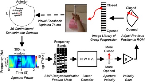
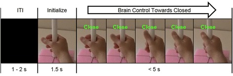
Methods: Utilizing magnetoencephalography's (MEG) high temporal and spatial resolution, we recorded sensorimotor rhythms (SMR) that were modulated by grasp or rest intentions. SMR modulation controlled the grasp aperture of a stop motion video of a human hand. The displayed hand grasp position was driven incrementally towards a closed or opened state and subjects were required to hold the targeted position for a time that was adjusted to change the task difficulty.

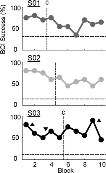
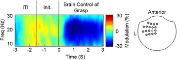
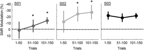
Results: We demonstrated that three individuals with complete hand paralysis due to spinal cord injury (SCI) were able to maintain brain-control of closing and opening a virtual hand with an average of 63 % success which was significantly above the average chance rate of 19 %. This level of performance was achieved without pre-training and less than 4 min of calibration. In addition, successful grasp targets were reached in 1.96 ± 0.15 s. Subjects performed 200 brain-controlled trials in approximately 30 min excluding breaks. Two of the three participants showed a significant improvement in SMR indicating that they had learned to change their brain activity within a single session of NF.

Conclusions: This study demonstrated the utility of a MEG-based BCI system to provide realistic, efficient, and focused NF to individuals with paralysis with the goal of using NF to induce neuroplasticity.


Figures

Fig. 1
Fig. 1
Schematic of the BCI used to translate SMR into proportional control of grasping. Beginning in the upper left, first, the power spectrum of data recorded from 36 sensorimotor MEG sensors (shown on a top-down view of the MEG helmet) are computed using 300 ms sliding windows. A mask is applied to these features to remove any components that did not exhibit desynchronization during calibration. Then a linear decoder applies weights (W) to the neural signal (N) to compute a hand velocity value (V H). The velocity output from the decoder is scaled (g) to ensure movement speeds are appropriate for the task. The previous hand position (an image from the video sequence) is then updated more closed or more opened within the ROM based on the scaled velocity command. The picture representing the desired aperture is chosen from 25 possible images. A progressive change in the images appeared to participants as a grasping movie with a 76 ms refresh rate
Fig. 2
Fig. 2
Trial timing. Participants proportionally controlled the hand to an opened or closed target-state during the brain-control phase. A stop motion video of grasping was progressed opened or closed based on brain activity. The full ROM spanned 25 frames of a stop-motion sequence (only 5 shown here). Trials were considered successful if the hand was held within 10 % of the target aperture for the given hold time (minimum of 500 ms)
Fig. 3
Fig. 3
BCI performance across blocks. Mean success rate for each block of 20 trials including 15 grasp and five rest trials. Horizontal dashed lines indicate individual subject chance levels computed with bootstrapping. Vertical dashed lines indicate when breaks happened. A “c” indicates that the decoder was recalibrated during the break. Up arrows indicate that the difficultly was increased by increasing the required hold time from 500 ms to 700 ms. Down arrows indicate the difficulty was decreased to a 500 ms hold time
Fig. 4
Fig. 4
Example signals during brain control of grasp. Average SMR modulation across 150 brain-controlled grasp trials in one sensorimotor sensor for subject S03. This sensor is highlighted in red on a top-down view of the MEG helmet on the right of this figure. At time zero the participant is cued to close the virtual hand by decreasing their SMR, i.e. desynchronization shown as blue. Trials began after an ITI, followed by a hand initialization stage. Modulation is the percent change relative to the SMR activity during the ITI
Fig. 5
Fig. 5
Improvement in SMR modulation across sessions. S01 and S02 show a significant improvement in the ability to modulate SMR compared to their first 50 trials, indicated by the * (p < 0.05; corrected for multiple comparisons). Error bars are the standard deviation across trials within each session-segment
Fig. 6
Fig. 6
Topography of SMR during the NF session. Changes in SMR modulation across the whole head during the beginning (trials 1–50), middle (trials 50–100), and end (trials 100–150) of NF training. Darker blue indicates stronger desynchronization during BCI grasp control. The location of the sensors used for NF are outlined in dotted lines on a top-down view of the MEG helmet (same as previous figures)

References

    1. Wilson TW, Fleischer A, Archer D, Hayasaka S, Sawaki L. Oscillatory MEG motor activity reflects therapy-related plasticity in stroke patients. Neurorehabil Neural Repair. 2011;25:188–93. doi: 10.1177/1545968310378511. - DOI - PubMed
    1. Schaechter JD. Motor rehabilitation and brain plasticity after hemiparetic stroke. Prog Neurobiol. 2004;73:61–72. doi: 10.1016/j.pneurobio.2004.04.001. - DOI - PubMed
    1. Tecchio F, Zappasodi F, Tombini M, Oliviero A, Pasqualetti P, Vernieri F, et al. Brain plasticity in recovery from stroke: an MEG assessment. Neuroimage. 2006;32:1326–34. doi: 10.1016/j.neuroimage.2006.05.004. - DOI - PubMed
    1. Jurkiewicz MT, Mikulis DJ, McIlroy WE, Fehlings MG, Verrier MC. Sensorimotor cortical plasticity during recovery following spinal cord injury: a longitudinal fMRI study. Neurorehabil Neural Repair. 2007;21:527–38. doi: 10.1177/1545968307301872. - DOI - PubMed
    1. Kokotilo KJ, Eng J, Curt A, Boyd LA. Reorganization and preservation of motor control of the brain in spinal cord injury: a systematic review. J Neurotrauma. 2009;26:2113–26. doi: 10.1089/neu.2008.0688. - DOI - PMC - PubMed

Publication types