. 2015 Nov 1;195(9):4514-23.
doi: 10.4049/jimmunol.1500668. Epub 2015 Sep 21.

Granzyme B-Mediated Activation-Induced Death of CD4+ T Cells Inhibits Murine Acute Graft-versus-Host Disease

Affiliations

Granzyme B-Mediated Activation-Induced Death of CD4+ T Cells Inhibits Murine Acute Graft-versus-Host Disease

Wei Du et al. J Immunol. .

Abstract

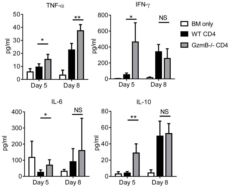
Granzyme B (GzmB) has previously been shown to be critical for CD8(+) T cell-mediated graft-versus-host disease (GVHD) but dispensable for GVHD mediated by CD4(+) T cells. However, previous studies used high doses of CD4(+) T cells in MHC-mismatched models that caused rapid and lethal GVHD. Because of the hyperacute lethality, it is possible that the role of GzmB was concealed by the system. Therefore, in this study, we have titrated down the T cell dose to precisely determine the contribution of GzmB in GVHD mediated by CD4(+)CD25(-) T cells. Surprisingly, we have found that GzmB(-/-)CD4(+)CD25(-) T cells cause more severe GVHD compared with wild-type CD4(+)CD25(-) T cells in both MHC-matched and mismatched models. Mechanistic analyses reveal that although GzmB does not affect donor T cell engraftment, proliferation or tissue-specific migration, GzmB(-/-) CD4(+)CD25(-) T cells exhibit significantly enhanced expansion because of GzmB-mediated activation-induced cell death of wild-type CD4(+)CD25(-) T cells. As a result of enhanced expansion, GzmB(-/-) T cells produced higher amounts of proinflammatory cytokines (e.g., TNF-α and IFN-γ) that may contribute to the exacerbated GVHD. These results reveal that GzmB diminishes the ability of CD4(+) T cells to cause acute GVHD, which contradicts its established role in CD8(+) T cells. The differential roles suggest that targeting GzmB in selected T cell subsets may provide a strategy to control GVHD.


Conflict of interest statement

Disclosure of Conflict of Interest

The authors have no potential conflict of interest to disclose.

Figures

Figure 1
Figure 1. GzmB is activated in donor CD4+CD25 T cells after allo-HCT
BALB/c (H-2d) host mice were lethally irradiated on day −1. On day 0, host mice were injected with 2×10⁶ TCD-BM cells alone or combined with 2×10⁴ WT or GzmB−/− CD4+CD25 T cells purified from C57BL/6 (H-2b) donor mice. On day 8, total cells harvested from the host spleens were analyzed with flow cytometry to examine GzmB expression in donor-derived T cells. Purified pre-HCT naive CD4+CD25 T cells were also examined as controls. (A) Representative dot plots are gated on donor-derived T cells (H-2Kb+CD3+CD4+). (B) Summary data of the percentages of donor CD4+CD25 T cells that are GzmB+ in the host spleens are shown with each point representing an individual mouse, with 9 mice assessed in each group.
Figure 2
Figure 2. GzmB−/− CD4+CD25 T cells cause more severe GVHD than WT CD4+CD25 T cells
BALB/c (H-2d) host mice were lethally irradiated on day −1. On day 0, mice were injected with 2×10⁶ TCD-BM alone or combined with 5×10⁴ (A) or 2×10⁴ (B) WT or GzmB−/− CD4+CD25 T cells purified from C57BL/6 (H-2b) donor mice and survival was monitored. (C) Allo-HCT was performed as described in (B) except that 2×10⁶ RAG1−/− BM cells were used instead of WT TCD-BM cells. (D) Shown are clinical GVHD scores for host mice on day 8 after allo-HCT in the experiment as described in (C). (E) C57BL/6 (H-2b) host mice were lethally irradiated on day −1. On day 0, host mice were injected with 7×10⁶ BM cells alone or combined with 4×10⁶ WT or GzmB−/− CD4+CD25 T cells isolated from 129/SvJ (H-2b) donor mice and survival was monitored. (F) Shown are clinical GVHD scores for host mice in the experiment described in (E). Summary survival results in each panel of (A,B,C,E) are combined from two independent experiments with similar results. Shown are Kaplan-Meier survival curves with Log-rank (Mantel-Cox) tests. Two-tailed t-tests were performed in (D) and Two-way-ANOVA in (F) to determine statistically significant differences.
Figure 3
Figure 3. GzmB−/− CD4+CD25 T cells cause more severe colon damage and bleeding
(A) BALB/c host mice were lethally irradiated on day −1. On day 0, mice were injected with 2×10⁶ TCD-BM cells alone or combined with 5×10⁴ WT or GzmB−/− CD4+CD25 T cells isolated from C57BL/6 donor mice. On day 8, host mice were dissected to examine GVHD damage. (B) C57BL/6 (H-2b) host mice were lethally irradiated on day −1. On day 0, host mice were injected with 7×10⁶ BM cells alone or combined with 4×10⁶ WT or GzmB−/− CD4+CD25 T cells isolated from 129/SvJ (H-2b) donor mice, On day 25, host mice were dissected to examine GVHD damage. Liver and colon histopathological GVHD scores were acquired as described the methods. (C) Representative image of intraluminal bleeding as indicated by a black arrow pointing at a colon segment that shows internal bleeding (upper panel). Representative images of intramural bleeding with black arrows pointing at areas of trapped red blood cells within the colon wall (lower panel). (D) Summary of the occurrence of observed colon bleeding in the host mice. Shown are summary results combined from two independent experiments with similar results.
Figure 4
Figure 4. GzmB−/− CD4+CD25 T cells caused more severe GI tract permeability
(A) BALB/c host mice were lethally irradiated with on day −1. On Day 0, mice were injected with 2×10⁶ TCD-BM cells alone or combined with 2×10⁴ WT or GzmB−/− CD4+CD25 T cells isolated from C57BL/6 (H-2b) donor mice. (B,C,D) C57BL/6 (H-2b) host mice were lethally irradiated on day −1. On day 0, mice were injected with 7×10⁶ BM cells alone or combined with 4×10⁶ WT or GzmB−/− CD4+CD25 T cells isolated from 129/SvJ (H-2b) donor mice. On the indicated time after allo-HCT, FITC-dextran GI tract permeability assay was performed as described in the methods with serum samples collected via eye bleeding. Summary data are shown with 7–12 mice assessed in each group at each time point. Two-tailed t-tests were performed to determine statistically significant differences.
Figure 5
Figure 5. GzmB−/− CD4+CD25 T cells exhibit enhanced expansion compared to WT cells
BALB/c host mice were lethally irradiated on day −1. On day 0, mice were injected with 2×10⁶ TCD-BM cells alone or combined with 5×10⁴ WT or GzmB−/− CD4+CD25 T cells isolated from C57BL/6 donor mice. (A) On day 5 after allo-HCT, splenocytes were harvested from the host mice and analyzed by flow cytometry. Representative flow plots showing CD3 and CD4 staining of live donor cells are gated on H-2Kb+Live/Dead cells. Summary data show the absolute total numbers of live donor T cells acquired by multiplying the percentages of H-2Kb+Live/Dead CD3+CD4+ cells to total cell numbers harvested from the host spleens (B), large intestines (C) and MLN (D). Data are shown as mean ± SD, with 4–6 mice assessed in each group at each time point. Two-tailed t-tests were performed to determine statistically significant differences.
Figure 6
Figure 6. GzmB is involved in activation-induced cell death of donor CD4+CD25 T cells
BALB/c host mice were lethally irradiated on day −1. On day 0, mice were injected with 2×10⁶ TCD-BM cells alone or combined with 3×10⁵ WT or GzmB−/− CD4+CD25 T cells isolated from C57BL/6 donor mice. On day 5 after allo-HCT, total cells harvested from the host spleens and MLN were assessed by Annexin V and Live/Dead dye staining to measure cell death. (A) Representative dot plots are gated on donor T cells (H-2Kb+CD3+CD4+) with naïve healthy donor mice serving as negative controls to show background cell death. (B) The percentages of donor T cells from the spleen (left panel) and MLN (right panel) that are Annexin V+ are shown, with 5 mice in each group. Background cell death indicated by the negative control was subtracted from each sample to show cell death induced specifically by allogeneic activation. Shown are representative data from one out of five independent experiments with similar results. Activated caspase 3 (C) and ROS (D) were stained to assess cell death. Shown are representative data from one out of three independent experiments with similar results. Two-tailed t-tests were performed to determine statistically significant differences.
Figure 7
Figure 7. GzmB−/− CD4+CD25 T cells induce higher amounts of proinflammatory cytokines
BALB/c (H-2d) host mice were lethally irradiated on day −1. On day 0, mice were injected with 2×10⁶ TCD-BM cells alone or combined with 5×10⁴ WT or GzmB−/− CD4+CD25 T cells isolated from C57BL/6 (H-2b) donor mice. Serum samples were collected via eye bleeding on day 5 and day 8 after allo-HCT. Serum cytokines levels were measured by luminex assays. Data are shown as mean ± SD, with 4–5 mice assessed in each group at each time point. Two-tailed t-tests were performed to determine statistically significant differences (*P<0.05; **P<0.01).

References

    1. Shlomchik WD. Graft-versus-host disease. Nat Rev Immunol. 2007;7:340–352. - PubMed
    1. Copelan EA. Hematopoietic stem-cell transplantation. N Engl J Med. 2006;354:1813–1826. - PubMed
    1. Welniak LA, Blazar BR, Murphy WJ. Immunobiology of allogeneic hematopoietic stem cell transplantation. Annu Rev Immunol. 2007;25:139–170. - PubMed
    1. Vogelsang GB, Lee L, Bensen-Kennedy DM. Pathogenesis and treatment of graft-versus-host disease after bone marrow transplant. Annu Rev Med. 2003;54:29–52. - PubMed
    1. Blazar BR, Murphy WJ, Abedi M. Advances in graft-versus-host disease biology and therapy. Nature reviews Immunology. 2012;12:443–458. - PMC - PubMed

Publication types

MeSH terms