. 2016 Feb;64(2):112-24.
doi: 10.1369/0022155415610792. Epub 2015 Sep 21.

Optimization of Single- and Dual-Color Immunofluorescence Protocols for Formalin-Fixed, Paraffin-Embedded Archival Tissues

Affiliations

Optimization of Single- and Dual-Color Immunofluorescence Protocols for Formalin-Fixed, Paraffin-Embedded Archival Tissues

Junko Kajimura et al. J Histochem Cytochem. 2016 Feb.

Abstract

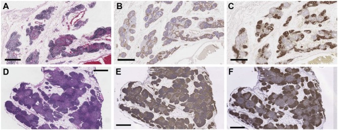
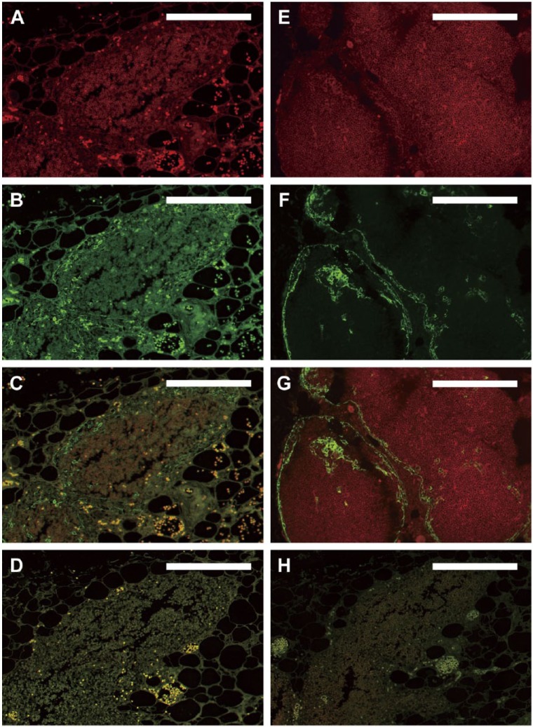
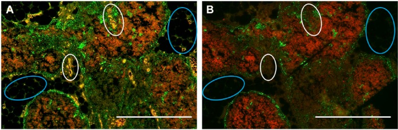
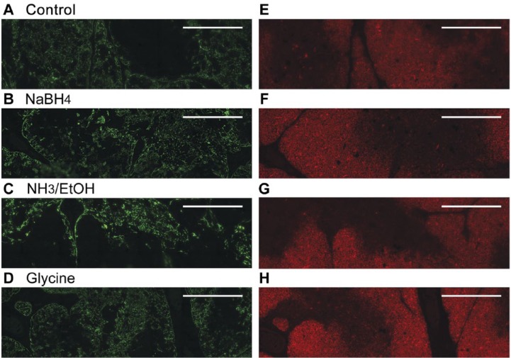
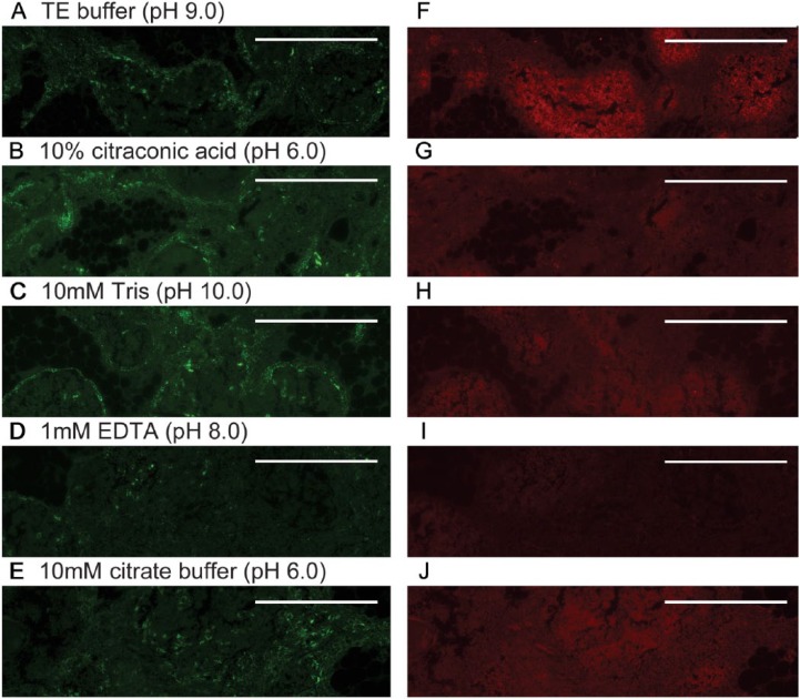
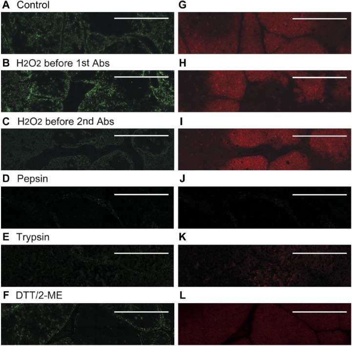
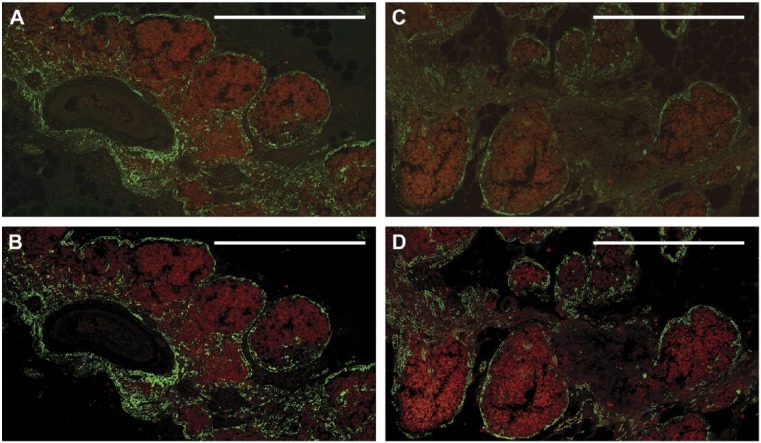
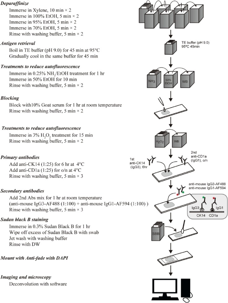
Performance of immunofluorescence staining on archival formalin-fixed paraffin-embedded human tissues is generally not considered to be feasible, primarily due to problems with tissue quality and autofluorescence. We report the development and application of procedures that allowed for the study of a unique archive of thymus tissues derived from autopsies of individuals exposed to atomic bomb radiation in Hiroshima, Japan in 1945. Multiple independent treatments were used to minimize autofluorescence and maximize fluorescent antibody signals. Treatments with NH3/EtOH and Sudan Black B were particularly useful in decreasing autofluorescent moieties present in the tissue. Deconvolution microscopy was used to further enhance the signal-to-noise ratios. Together, these techniques provide high-quality single- and dual-color fluorescent images with low background and high contrast from paraffin blocks of thymus tissue that were prepared up to 60 years ago. The resulting high-quality images allow the application of a variety of image analyses to thymus tissues that previously were not accessible. Whereas the procedures presented remain to be tested for other tissue types and archival conditions, the approach described may facilitate greater utilization of older paraffin block archives for modern immunofluorescence studies.

Keywords: archival tissue; image analysis; immunofluorescence; thymus; tissue processing.


Conflict of interest statement

Competing Interests: The authors declared no potential conflicts of interest with respect to the research, authorship, and/or publication of this article.

Figures

Figure 1.
Figure 1.
H&E and bright-field immunohistochemical staining of archival thymus tissues. (A–C) RERF thymus tissues were fixed in unbuffered formalin, processed into paraffin blocks, and stored for ~50 years under initially uncontrolled conditions before they were stained with (A) H&E, or with (B) anti-CK14, or (C) anti-CD1a antibodies in immunoperoxidase assays, as described in the Materials & Methods. The results closely modeled those obtained (D–F) using thymus tissues that were fixed with neutral-buffered formalin and stored under climate-controlled conditions for 20–25 years before they were stained with (D) H&E, (E) anti-CK14, or anti-CD1a (F) antibodies using identical methods. Scale, 1 mm.
Figure 2.
Figure 2.
Immunofluorescence using standard protocols. Dual-color immunofluorescence images were derived from older RERF archival thymus tissues fixed in unbuffered formalin and stored under initially uncontrolled conditions (A–D, H). (A–C) Fluorescence following reaction with anti-CD1a antibody (red, A), anti-CK14 antibody (green, B), and the merged images (C) on a single slide, in the absence of autofluorescence-reducing techniques. A separate section of the same tissue that was deparaffinized but not reacted with antibodies is shown in (D) to document autofluorescence in the absence of background antibody staining. (A–D) High autofluorescence and poor signal-to-noise ratio initially observed with the older archival tissues that prevented definitive identification of cortical thymocytes in (A) and thymic epithelium in (B). For comparison, thymus tissues fixed with neutral-buffered formalin and stored under climate-controlled conditions for 25 years before staining (E–G) show optimal dual-color immunofluorescence images under the same staining conditions used for panels (A–C). Strong specific staining of cortical thymocytes (red, E) and thymic epithelial cells (green, F; merged images in G) is observed in these tissues without the need for additional autofluorescence-reducing techniques. The staining pattern shown in (E–G) thus served as a gold standard for determining optimal IF staining of the older archival tissues from RERF throughout the remainder of the study. (H) Same tissue as in (A–D) with omission of primary antibodies but with application of NH3/EtOH, H2O2, and Sudan Black B treatments, as described in the text. Scale, 0.2 mm.
Figure 3.
Figure 3.
Sudan black staining quenches natural autofluorescence. (A) White circles indicate foci of red blood cells (RBCs) and blue circles indicate elastin fibers that generate strong autofluorescence and obscure the signal resulting from bound anti-CK14 antibody in the tissue shown in Figure 2. A serial section with Sudan black staining applied after antibody staining but before coverslipping (B) shows that this treatment decreases autofluorescence of these natural components to almost non-detectable levels. Scale, 0.5 mm.
Figure 4.
Figure 4.
Treatment with a mild reducing agent reduces autofluorescence without markedly decreasing antibody signals. Representative dual immunofluorescence images obtained with anti-CK14 antibody (green, A–D) or anti-CD1a antibody (red, E–H) are shown without (A, E) and with additional steps to reduce autofluorescence: NaBH4 (B, F), NH3/ethanol (C, G), or glycine (D, H). See Materials & Methods for details of treatments. The NH3/EtOH treatment decreased autofluorescence so that internal cortical areas now appear appropriately negative with anti-CK14 antibody (green, panel C) and medullary areas now appear appropriately negative with anti-CD1a antibody (red, panel G). Scale, 0.5 mm.
Figure 5.
Figure 5.
Methods to enhance antibody reactivity/signals. Representative dual immunofluorescence images obtained with anti-CK14 (green, A–E) or anti-CD1a antibody (red, F–J) are shown after heating at 95°C for 45 min in various buffers in an attempt to enhance antibody reactivity/signals. Buffers used were: TE buffer, pH 9.0 (A, F); 10% citraconic acid, pH 6.0 (B, G); 10 mM Tris, pH 10.0 (C, H); 1 mM EDTA, pH 8.0 (D, I); and 10 mM citrate buffer, pH 6.0 (E, J). All tissues shown were stained with 0.3% Sudan Black B to reduce autofluorescence, but did not receive NH3/EtOH or 3% H2O2 treatments. Scale, 0.5 mm.
Figure 6.
Figure 6.
Methods to enhance antibody reactivity/signals. Representative immunofluorescence images obtained with anti-CK14 antibody (A–F) or anti-CD1a antibody (G–L) are shown without (A, G) and with additional steps to enhance antibody reactivity/signals: H2O2 in methanol prior to primary antibody (B, H): H2O2 in methanol prior to secondary antibody (C, I); pepsin (D, J); trypsin (E, K); or DTT/2-ME (F, L). See Materials & Methods for details of treatments. Scale, 0.5 mm.
Figure 7.
Figure 7.
Application of deconvolution microscopy further reduces background and improves the signal-to-noise ratio in dual-color immunofluorescence. The two different thymus tissues shown were each fixed in unbuffered formalin and stored as described in the Materials & Methods for 52 years before staining with anti-CK14 (green) and anti-CD1a (red) antibodies. Treatments to reduce autofluorescence and enhance signal intensity incubation were NH3/EtOH, 3% H2O2, and 0.3% Sudan Black B, as described in the Materials & Methods. Prior to deconvolution, fluorescence signals are blurred and unclear (A, C). The addition of deconvolution microscopy (B, D) further improves the images, resulting in a sharper signal that enhances image quality. Signal intensities and clarity are now similar to those seen with the more recent tissues (compare with Fig. 2G), and allow for the differential localization of cell types. Scale, 0.5 mm.
Figure 8.
Figure 8.
Optimized protocol to minimize autofluorescence and maximize antibody signals. Shown is a summary of the protocol used to generate high-quality, dual fluorescence images from archival thymus tissues. Abbreviations: o/n, overnight; Abs, antibodies; DW, distilled water.

References

    1. Baschong W, Suetterlin R, Laeng RH. (2001). Control of autofluorescence of archival formaldehyde-fixed, paraffin-embedded tissue in confocal laser scanning microscopy (CLSM). J Histochem Cytochem 49:1565-1572. - PubMed
    1. Beisker W, Dolbeare F, Gray JW. (1987). An improved immunocytochemicalprocedure for high-sensitivity detection of incorporated bromodeoxyuridine. Cytometry 8:235-239. - PubMed
    1. Kiernan JA. (2008). Histological and Histochemical Methods: Theory and Practice. 4th edition, Bloxham: Scion.
    1. Oliveira VC, Carrara RC, Simoes DL, Saggioro FP, Carlotti CG, Covas DT, Neder L. (2010). Sudan Black B treatment reduced autofluorescence and improves resolution of in situ hybridization specific fluorescent signals of brain sections. Histol Histopathol 25:1017-1024. - PubMed
    1. Shi S-R, Shi Y, Taylor CR. (2011). Antigen Retrieval Immunohistochemistry: Review and future prospects in research and diagnosis over two decades. J Histochem Cytochem 59:13-32. - PMC - PubMed

Publication types

LinkOut - more resources