. 2015 Oct 6;112(40):12402-7.
doi: 10.1073/pnas.1511308112. Epub 2015 Sep 21.

Thermodynamics of protein destabilization in live cells

Affiliations

Thermodynamics of protein destabilization in live cells

Jens Danielsson et al. Proc Natl Acad Sci U S A. .

Abstract

Although protein folding and stability have been well explored under simplified conditions in vitro, it is yet unclear how these basic self-organization events are modulated by the crowded interior of live cells. To find out, we use here in-cell NMR to follow at atomic resolution the thermal unfolding of a β-barrel protein inside mammalian and bacterial cells. Challenging the view from in vitro crowding effects, we find that the cells destabilize the protein at 37 °C but with a conspicuous twist: While the melting temperature goes down the cold unfolding moves into the physiological regime, coupled to an augmented heat-capacity change. The effect seems induced by transient, sequence-specific, interactions with the cellular components, acting preferentially on the unfolded ensemble. This points to a model where the in vivo influence on protein behavior is case specific, determined by the individual protein's interplay with the functionally optimized "interaction landscape" of the cellular interior.

Keywords: NMR; crowding; in vivo; protein stability; thermodynamics.


Conflict of interest statement

The authors declare no conflict of interest.

Figures

Fig. S1.
Fig. S1.
The structure of the native SOD1 dimer (PDB code 1HL5) and the loop regions removed by protein engineering. (A) In the native SOD1 dimer the long loops IV and VII adapt a compact and highly ordered structure around the active site, where loop IV also forms part of the dimer interface (green). The left-hand monomer is shown as accessible surface (1.4 Å probe radius) whereas the right-hand monomer is represented as a cartoon. Highlighted are the residues coordinating the active-site Cu1+/2+ and Zn2+ ions and the C57–C146 disulfide linkage between loop IV and the central β-barrel. (B) Removal of loops IV and VII from apoSOD1 reduces the dimer interface as well as the metal binding moieties and leads to soluble apoSOD1barrel monomers (PDB code 4BCZ). The truncated loops IV and VII are highlighted in green and blue, respectively. (Adapted from ref. .)
Fig. S2.
Fig. S2.
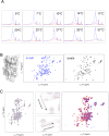
(A) Controls of internalization. One-dimensional 15N-HMQC spectra of SOD1I35A in A2780 cells (blue) and in supernatant (red) show no or small amounts of leakage. (B, Left) X-ray structure of SOD1barrel (PDB code 4BCZ), representing the β-barrel scaffold of the ALS-associated protein Cu/Zn superoxide dismutase 1 (66). The method yields here intracellular concentrations of 20–30 μM, matching those of human SOD1 in transgenic ALS mice (43) and in vitro aggregation studies (6). (B, Center and Right) HMQC spectra of SOD1barrel in mammalian cells (blue) and the subsequent cell lysate (black). (C, Left) Overlay of 1H-{15N}-HMQC spectra obtained at pH values ranging from 5.8 to 7.6. (C, Center) Close-ups of the most affected cross peaks. (C, Right) Overlay of the best-fit in vitro spectra (red) and the in-cell spectra (blue), used together with the rest of the data to estimate the pH inside the A2780 cells to 6.5 ± 0.1 (32).
Fig. 1.
Fig. 1.
In vitro benchmarking of SOD1barrel, poised at marginal thermodynamic stability by the mutation SOD1I35A. (A) HMQC spectra of SOD1barrel at 37 °C, showing uniformly folded protein. Inset shows the X-ray structure of SOD1barrel (PDB code 4BCZ), constituting the β-barrel scaffold of the parent ALS-associated protein Cu/Zn superoxide dismutase 1 (32). (B) Corresponding HMQC spectra of the mutant SOD1I35A (PDB code 4XCR), showing mixed population of folded (N) and unfolded (D) material. Quantification of the D/N equilibrium is from the cross-peak volumes of the C-terminal resonance Q153. (C) ΔGD-N vs. temperature profiles of SOD1barrel and SOD1I35A obtained from NMR thermal scans. The populations of D and N vs. temperature show melting a point Tm = 35.4 °C, i.e., ΔGD-N = 0 (Eq. 1), and cold unfolding at subzero temperature. The curved ΔGD-N profiles with stability maxima around room temperature are characteristic for naturally evolved proteins (58) and define a standard set of thermodynamic parameters with well-established structural meaning (Eq. 2).
Fig. S3.
Fig. S3.
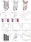
(A–C) The crystal structure of SOD1I35A (PDB code 4XCR) (red) overlaid with that of the SOD1barrel (PDB code 4BCZ) (gray), showing that the folded states of the two protein variants are largely the same. (D and E, Upper) In vitro HMQC spectra of marginally stable SOD1I35A and the fully folded SOD1barrel. (D and E, Lower) The electrostatic surfaces of the two proteins. Taken together, the data show that the structures and surface properties of SOD1I35A and SOD1barrel are very similar. (F) Chevron plots of SOD1I35A at 17 °C, 25 °C, and 37 °C. The lower temperatures show the signature of a two-state folder (28), whereas at 37 °C the midpoint is too low to observe any refolding of the protein. (G) The free-energy profile of SOD1I35A in A2780 cells (blue) and in vitro data with a simulated varying selective line broadening of the Q153 cross peak of the folded state, PF (red). (H) The factors of line broadening applied to PF to reproduce in-cell data (blue) compared with the factors measured from in-cell data (red) and the factors measured in high-viscosity in vitro samples (black). (I) Calculated maximum effect on ΔGD-N from low-temperature viscosity effects. The peak-volume determination can suffer from systematic errors due to line broadening at low temperatures, but the maximum effect is less than 250 J⋅mol−1. (J) The ratio of R2 for the folded and unfolded Q153 cross peak at 280 K, 290 K, and 310 K shows only moderate temperature dependence. (K) Overlay of HMQC spectra of the folded SOD1barrel and the fully unfolded double-mutant SOD1I35A/G93A. (L) The relative peak volumes of Q153 D (red) and N (black) as a function of temperature. The calculated population of N remains constant at 0.5 (blue), as expected for a system with only a small contribution from line broadening and relaxation bias.
Fig. S4.
Fig. S4.
(A) Free-energy profiles of SOD1barrel (blue) and SOD1I35A (green), derived from CD-melting data (Inset), compared with the free-energy profile of SOD1I35A determined by NMR (orange). (B) The pH dependence of Tm and ΔH of SOD1I35A used to determine ΔCp from the relationship ΔCp = δΔHTm. (C) In-cell sample stability vs. time. The 1D 1H-{15N}-HMQC spectra of SOD1I35A in A2780 cells at the start of the experiment (red) and after 2D acquisition (blue) are overall similar. (D–H) Controls of pH and salt effects. (D) Population folded SOD1I35A material determined from Q153 cross peaks as a function of pH. (E) Thermal melting point of SOD1I35A determined by CD as a function of pH. (F) Free-energy profiles determined from in-cell data directly (blue) and data skewing arising by offsetting the pH to, 6.25 (red) and 6.72 (black). (G) Free-energy profiles for SOD1I35A without added salt (orange) and in increasing amounts of NaCl, up to 300 mM. (H) The melting temperatures for SOD1I35A at the NaCl concentrations used in G.
Fig. 2.
Fig. 2.
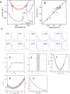
In-cell quantification of protein stability. (A) Schematic illustration of protein delivery by electroporation. The method yields intracellular concentrations of SOD1I35A = 20–30 μM, matching those of human SOD1 in transgenic ALS mice (43) and in vitro aggregation studies (6). (B) Two overlaid in-cell HMQC spectra of SOD1I35A showing that the protein is mainly folded at 17 °C (red) and fully unfolded already at 37 °C (blue). An advantage of this detection strategy is that the target protein is retained fully physiological and devoid of potentially interfering spectroscopic reporters. (C) ΔGD-N vs. temperature profiles based on quantification of the D/N equilibrium from the Q153 cross-peak volumes. The results show that both mammalian and bacterial cells substantially destabilize SOD1I35A, albeit in slightly different ways. A common feature is that the in-cell destabilization shifts both cold unfolding (TC) and melting temperatures (Tm) to the physiological regime (Table 1).
Fig. S5.
Fig. S5.
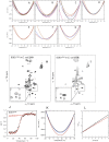
Free-energy profiles used for in vitro controls. The in vitro references (PBS) are shown in orange. (A) The effect of E. coli lysates on SOD1I35A stability is critically sensitive to lysate preparation. (B) Increasing amounts of the inert crowder Ficoll 70 increases SOD1I35A stability: 50 mg/mL (green), 100 mg/mL (red), 150 mg/mL (blue), 200 mg/mL (black), and 250 mg/mL (pink). (C) The corresponding effect of PEG400: 5% (brown), 10% (blue), 20% (black), and 30% (pink). (D) Crowding with BSA yields only moderate effects: 40 mg/mL (red), 80 mg/mL (blue), and 100 mg/mL (black). (E) The corresponding effect of 100 mg/mL holoSOD1dimer (red). (F) Crowding with TTHApwt: 50 mg/mL (blue) and 100 mg/mL (red). (G) Crowding with lysozyme yields marked destabilization even at low concentrations: 30 mg/mL (blue) and 50 mg/mL (red). (H and I) In-E. coli HMQC spectra of SOD1I35A at 290 K and 310 K show line broadening. Even so, the narrow cross peaks of the dynamic C-terminal Q153 can be used for accurate determination of the D and N populations. (J–L) Thermodynamic and kinetic similarity of SOD1I35A and reduced apoSOD1pwt. Thermal melting of the two SOD1 variants shows inseparable traces. (K) Comparison of free-energy profiles of SOD1I35A derived from NMR (black) and CD data (blue) and the free-energy profile of apoSOD1pwt derived from CD data (red). (L) SOD1I35A and reduced apoSOD1pwt show similar unfolding rates and similar linear urea dependence, suggesting similar folded structures (66).
Fig. 3.
Fig. 3.
Comparison of in vivo and in vitro data, showing that osmolytes yield stability changes opposite to those of the cells, whereas protein crowders yield the whole spectrum of effects, underlining the amino acid sequence dependence of the protein solute interactions. The solute concentrations of A2780 and E. coli cells show considerable variation in the literature (13), spanning the range of the error bars.
Fig. 4.
Fig. 4.
Coupled folding equilibrium describing the shift toward denatured material upon interaction with the cellular interior, as formalized in Eqs. 35. Both the denatured (D) and folded (N) species interact with the cellular molecules (m), but the interactions are stronger/more numerous for the structurally expanded and flexible D species. The increased heat capacity of unfolding (ΔCp) observed in the cellular compartment (Eqs. 1 and 2 and Fig. 2) is attributed to increased solvent-accessible surface area (dotted boundary) of the denatured ensemble (D cell), promoted by the transient association with neighboring macromolecules (m). Following Elcock’s estimate (44), SOD1I35A would at all times experience approximately five putative interaction partners in its immediate cellular environment. Associated thermodynamic parameters are in Table 1 and Table S2.

References

    1. Joerger AC, Fersht AR. Structural biology of the tumor suppressor p53. Annu Rev Biochem. 2008;77:557–582. - PubMed
    1. Bergemalm D, et al. Overloading of stable and exclusion of unstable human superoxide dismutase-1 variants in mitochondria of murine amyotrophic lateral sclerosis models. J Neurosci. 2006;26(16):4147–4154. - PMC - PubMed
    1. Luchinat E, et al. In-cell NMR reveals potential precursor of toxic species from SOD1 fALS mutants. Nat Commun. 2014;5:5502. - PubMed
    1. Byström R, Andersen PM, Gröbner G, Oliveberg M. SOD1 mutations targeting surface hydrogen bonds promote amyotrophic lateral sclerosis without reducing apo-state stability. J Biol Chem. 2010;285(25):19544–19552. - PMC - PubMed
    1. Ciryam P, Tartaglia GG, Morimoto RI, Dobson CM, Vendruscolo M. Widespread aggregation and neurodegenerative diseases are associated with supersaturated proteins. Cell Reports. 2013;5(3):781–790. - PMC - PubMed

Publication types

MeSH terms

Associated data