. 2015 Nov;21(11):1966-79.
doi: 10.1261/rna.053629.115. Epub 2015 Sep 21.

Extracellular vesicle-mediated transfer of processed and functional RNY5 RNA

Affiliations

Extracellular vesicle-mediated transfer of processed and functional RNY5 RNA

Sudipto K Chakrabortty et al. RNA. 2015 Nov.

Abstract

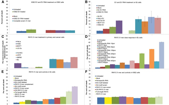
Extracellular vesicles (EVs) have been proposed as a means to promote intercellular communication. We show that when human primary cells are exposed to cancer cell EVs, rapid cell death of the primary cells is observed, while cancer cells treated with primary or cancer cell EVs do not display this response. The active agents that trigger cell death are 29- to 31-nucleotide (nt) or 22- to 23-nt processed fragments of an 83-nt primary transcript of the human RNY5 gene that are highly likely to be formed within the EVs. Primary cells treated with either cancer cell EVs, deproteinized total RNA from either primary or cancer cell EVs, or synthetic versions of 31- and 23-nt fragments trigger rapid cell death in a dose-dependent manner. The transfer of processed RNY5 fragments through EVs may reflect a novel strategy used by cancer cells toward the establishment of a favorable microenvironment for their proliferation and invasion.

Keywords: RNY5; cancer microenvironment; exosomes; extracellular vesicles.


Figures

FIGURE 1.
FIGURE 1.
Validation of purification of extracellular vesicles (EVs). (A) Transmission electron microscopy image of K562 EVs after negative staining shows classic cup-shaped vesicles that are on average smaller than 200 nm. (B) Immuno-electron microscopy image of purified EVs labeled with anti-CD81 (mouse mAb) and detected by goat anti-mouse IgG secondary conjugated with 5 nm gold. Dark spots on the image are the electron dense gold elements conjugate to IgG secondary antibody. (C) Bioanalyzer RNA profile (RNA Pico-chip) of untreated EVs (red), RNA profile of EVs treated with RNase (green), and RNA profile of EVs treated with detergent and RNase (blue). (X-axis) Nucleotide lengths; (y-axis) fluorescent units. (D) Western blot analysis of proteins from K562 EVs and whole cell. Proteins selected for detection were previously identified to be enriched in EV or whole cell. (EV enriched) ALIX (PDCD6IP gene), CD71 (TfR1 gene), TSG101 (TSG101 gene). (Whole cell) PDI (PDI gene), FIBRILLARIN (FBL gene), PROHIBITIN (PHB gene).
FIGURE 2.
FIGURE 2.
Pie charts representing the relative abundance of families of RNA within BJ whole cell (A), K562 whole cell (B), BJ EV (C), and K562 EV (D). The group labeled as “Others” in the pie charts is representative of reads derived from several Gencode annotation categories such as pseudogenes, antisense intronic, mitochondrial t-RNA, vault RNA, immunoglobulin genes, etc.
FIGURE 3.
FIGURE 3.
Fragmentation patterns of RNY5. (A) Full-length RNY5 structure. The structure was drawn using Mfold according to data from van Gelder et al. (1994). Bold line indicates the 5′ 31-nt processed product and the 8-nt motif is highlighted. (B) Graph depicting the most frequent (>1000 reads per million) start and stop positions of reads mapping to the human RNY5 gene. The most frequent start positions marked as the 5′ start position of the RNY5 annotation and position 52 of the annotation. The most frequent stop positions are 23, 29, and 31 for the reads, which start at the 5′ end of the RNY5 gene, and position 83 which has reads starting at 52 and also some reads that start at position 1. (C) Northern blot of RNY5 RNA purified from K562, BJ cells, and EVs. Synthetic versions of Y5 processing products were used as size markers. RNA was detected by a probe complementary to the 5′ 31-nt processed product. (w) Whole-cell RNA, (e) EV RNA. (D,E) In vitro processing of RNY5. Synthetic full-length RNY5 was incubated for 30 min at 37° with 0, 2, 4, or 8 µg of K562 whole cell (D) or EV (E) protein extract. Samples containing only the extracts and treated identically were used to control for the existence of Y5 RNA in protein extracts. Detection was done as in C. Note that 23- and 31-nt size markers are not equimolar. (F) In vitro processing of Y5 5′ 31-mer variants. Wild-type (WT), scrambled (scram.), and 8-nt motif scrambled (motif scram.) versions of the Y5 5′ 31-mer were radioactively end-labeled and incubated with K562 EV protein extract for 2 h at 37°.
FIGURE 4.
FIGURE 4.
Quantification of cell death by flow cytometry. YO-PRO-1 and Hoechst33342 dyes were used for quantification of cell death. Y-axis indicates the percent of cell death indicated by YO-PRO-1 and Hoechst33342 double positive cells. The mean of duplicates is presented with error bars indicating variation from mean. (A) Levels of cell death in K562 cells when treated with EVs and EV RNA. Y-axis indicates percent cell death observed. The following treatments are presented: (Blue) untreated: K562 cells without any treatment; (green) K562 EV treated: K562 cells incubated with K562 EVs; (red) mock: K562 cells with Lipofectamine treated only (no RNA); (purple) K562 EV RNA treated: K562 cells treated with K562 EV RNA; (turquoise) complete scram 31-mer: k562 cells treated with 31-nt scrambled sequence. (B) Levels of cell death in BJ cells when treated with EVs and EV RNA. Y-axis indicates percent cell death observed. The following treatments are presented: (Blue) untreated: BJ cells without any treatment; (red) mock: BJ cells with lipofectamine treated only (no RNA); (green) BJ EV RNA: BJ cells transfected with BJ EV RNA; (purple) K562 EV RNA: BJ cells treated with K562 EV RNA; (light green) BJ EV: BJ cells incubated with BJ EVs; (turquoise) HeLa EV: BJ cells incubated with HeLa EVs; (orange) U2-Os EV: BJ cells incubated with U2-Os EVs; (light blue) MCF7 EV: BJ cells incubated with MCF7 EVs; (mauve) K562 EV: BJ cells incubated with K562 EVs. (C) Generality of RNY5 31-mer-induced cell-death phenotype. Bars indicate the net increase in cell death normalized to levels of cell death from mock treatment in each cell type. Four cancer cell lines including (blue) K562 (chronic myelogenous leukemia); (purple) HeLa (cervical adenocarcinoma); (red) MCF7 (breast adenocarcinoma); (green) U2-Os (osteosarcoma), and four primary cells including (light mauve) BJ (normal skin fibroblasts); (turquoise) HUVEC (normal human umbilical vein endothelial cell); (light blue) IMR90 (normal human lung fibroblasts); and (orange) HFFF (normal human fetal foreskin fibroblasts) were transfected with RNY5 31-mer. One hundred picomoles of RNY5 was used for each transfection, except (orange) HFFF, where 200 pmol of RNY5 31-mer was used. (D) Dose response curve of RNY5 31-mer-induced cell-death phenotype in BJ cells. The bars represent the percent of cell death when BJ cells are treated with increasing dose (10, 50, 100, 200, 300, and 400 pmol) of RNY5 31-mer or nonspecific RNA. AllStars negative control RNA (Qiagen) was used as a nonspecific RNA control. The levels of cell death in Untreated or Mock treated (Lipofectamine only) BJ cells are also indicated. (E) Levels of cell death in BJ cells from 100 pmol of synthetic RNA oligonucleotides transfection. Y-axis indicates the percent cell death. The synthetic RNA oligonucleotides used for transfection are as follows: (Blue) untreated: BJ cells without any treatment; (yellow) mock: BJ cells with Lipofectamine treated only (no RNA); (green) nonspecific RNA: nonspecific RNA control (AllStars negative control siRNA); (purple) 8-nt motif deleted: RNY5 sequence with nucleotides 14–21 motif deleted; (turquoise) RNY5 31-mer complement: 31-nt RNY5 3′ side fragment; (light orange) 8-nt motif scrambled: RNY5 31-mer sequence with nucleotides 14–21 scrambled; (blue) RNY5 31-mer scram: 31-nt completely scrambled sequence; (orange) DS RNY5 31-mer, double-stranded RNY5 31-mer duplex; (green) full-length RNY5: RNY5 83-mer full-length sequence; (light green) RNY5 31-mer: 5′ RNY5 31-nt fragment; (light purple) RNY5 23-mer: 5′ side RNY5 23-nt fragment. (F) Levels of cell death observed in K562 cells from 100 pmol of synthetic RNA oligonucleotides transfection. Y-axis indicates percent cell death. The synthetic RNA oligonucleotides used for transfection are as follows: (Blue) untreated: K562 cells without any treatment; (yellow) mock: K562 cells with Lipofectamine treated only (no RNA); (green) nonspecific RNA: nonspecific RNA control (AllStars negative control siRNA); (purple) 8-nt motif deleted: RNY5 sequence with nucleotides 14–21 motif deleted; (orange) 8-nt motif scrambled: RNY5 31-mer sequence with nucleotides 14–21 scrambled; (blue) RNY5 31-mer scram: 31-nt completely scrambled sequence; (red) DS RNY5: double stranded; (green) full-length RNY5 83-mer; (light green) RNY5 31-mer: 5′ RNY5 31-nt fragment.
FIGURE 5.
FIGURE 5.
The part of the TGF-β pathway depicted from KEGG pathways, where we observe similar changes in transcript levels of genes between both types of treatments (K562 EV and the 32-nt synthetic RNA) in both BJ and HUVEC cells. The pathway highlights a common response of transcript levels of most genes in this part of the pathway.

References

    1. Abusamra AJ, Zhong Z, Zheng X, Li M, Ichim TE, Chin JL, Min WP. 2005. Tumor exosomes expressing Fas ligand mediate CD8+ T-cell apoptosis. Blood Cells Mol Dis 35: 169–173. - PubMed
    1. Anders S, Huber W. 2010. Differential expression analysis for sequence count data. Genome Biol 11: R106. - PMC - PubMed
    1. Anders S, Pyl PT, Huber W. 2015. HTSeq–a Python framework to work with high-throughput sequencing data. Bioinformatics 31: 166–169. - PMC - PubMed
    1. Bidard FC, Pierga JY, Vincent-Salomon A, Poupon MF. 2008. A “class action” against the microenvironment: do cancer cells cooperate in metastasis? Cancer Metastasis Rev 27: 5–10. - PMC - PubMed
    1. Chen X, Smith JD, Shi H, Yang DD, Flavell RA, Wolin SL. 2003. The Ro autoantigen binds misfolded U2 small nuclear RNAs and assists mammalian cell survival after UV irradiation. Curr Biol 13: 2206–2211. - PubMed

Publication types