. 2015 Sep 9:9:5169-83.
doi: 10.2147/DDDT.S81578. eCollection 2015.

Regulation of bone formation by baicalein via the mTORC1 pathway

Affiliations

Regulation of bone formation by baicalein via the mTORC1 pathway

Sheng-fa Li et al. Drug Des Devel Ther. .

Abstract

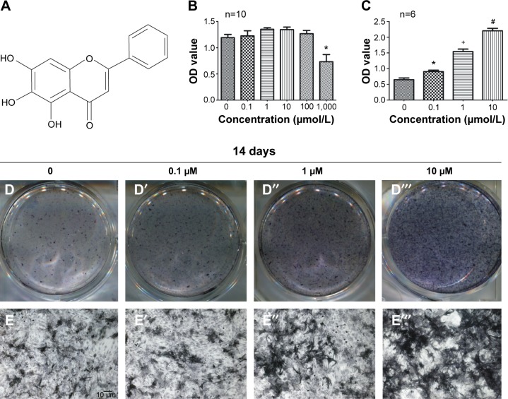
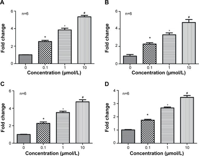
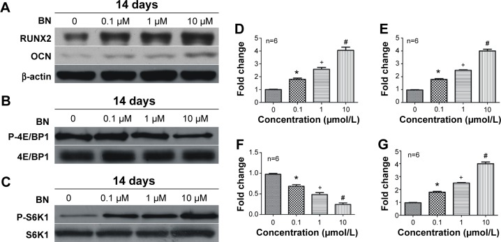
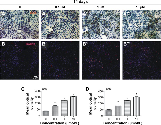
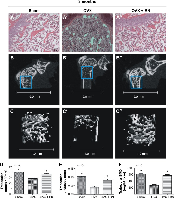
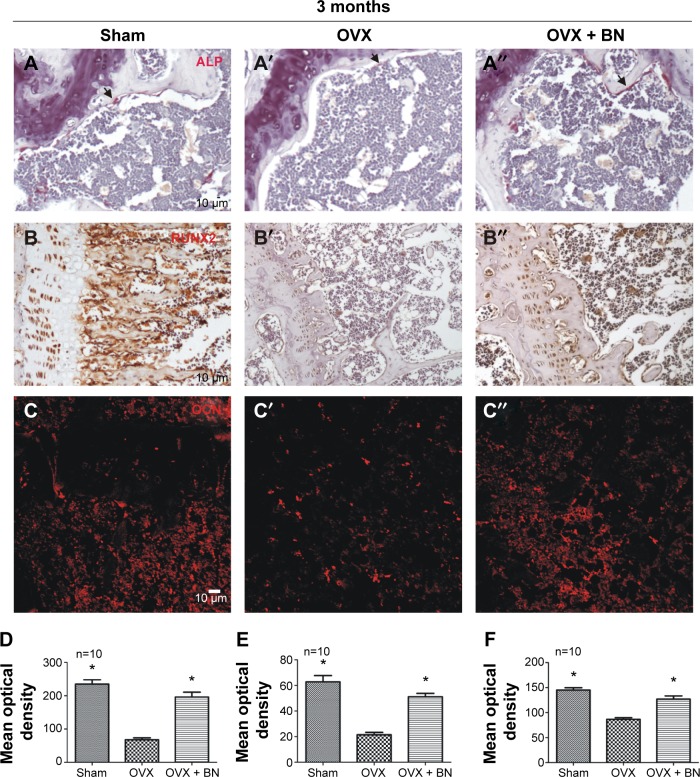
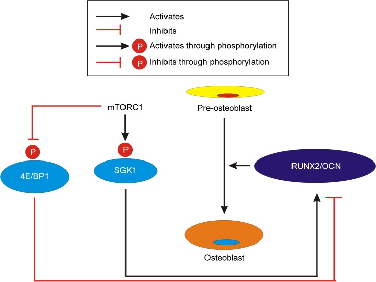
Osteoporosis is a systemic skeletal disease that is characterized by low bone density and microarchitectural deterioration of bone tissue. The increasing prevalence of osteoporosis has attracted much attention. In this study, MC3T3-E1 pre-osteoblasts were treated with the natural compound, baicalein (0.1 μmol/L, 1 μmol/L, 10 μmol/L), to stimulate differentiation over a 14-day period. In addition, a canonical ovariectomized (OVX) mouse model was used to investigate the effect of 3-month baicalein treatment (10 mg/kg per day) in preventing postmenopausal osteoporosis. In vitro, we found that baicalein induced activation of alkaline phosphatase, stimulated the mammalian target of rapamycin complex 1 (mTORC1) signaling pathway, and induced expression of osteoblast differentiation markers, ie, osteocalcin, osterix, collagen Iα1, and runt-related transcription factor 2 (RUNX2), in osteoblasts. In vivo, several bone parameters, including trabecular thickness, trabecular bone mineral density, and trabecular number, in the distal femoral metaphysis were significantly increased in OVX mice treated intragastrically with baicalein for 3 months compared with OVX mice that were not treated with baicalein. We also found that expression of osteocalcin and RUNX2 was decreased in primary ossified tissue from the OVX group, and baicalein increased the levels of osteocalcin and RUNX2 in OVX mice. These data suggest that baicalein can stimulate MC3T3-E1 cells to differentiate into osteoblasts via activation of the mTORC1 signaling pathway, which includes protein kinases and transcription factors such as P-4E/BP1 and P-S6K1.

Keywords: mTOR; menopause; osteoblast; osteoporosis.


Figures

Figure 1
Figure 1
MC3T3-E1 cells increased osteogenesis in the presence of BN. Notes: (A) Molecular structure of BN. (B) CCK-8 assays showed that BN did not significantly affect the growth of MC3T3-E1 cells at the concentrations used (0.1 μmol/L, 1 μmol/L, 10 μmol/L, 100 μmol/L, and 1,000 μmol/L) after treatment for 14 days, and BN at concentrations of 0.1–100 μM did not significantly affect cell growth. (C) The ALP activity assay showed that BN increased intracellular ALP expression during osteoblast differentiation in cultured MC3T3-E1 cells. (D) ALP staining indicates that treatment with 0.1–10 μM BN increased ALP activity in MC3T3-E1 cells in osteogenic differentiation medium for 14 days. (E) Micrograph of ALP staining indicates that the expression of ALP shows dose-dependent with 0.1–10 μM BN treatment. *P<0.05 versus group without BN; +P<0.05 versus group with 0.1 μM BN; #P<0.05 versus group with 1 μM BN. Columns represent the mean ± standard error from six wells per group (B, C). Abbreviations: ALP, alkaline phosphatase; BN, baicalein; CCK-8, Cell Counting Kit-8; OD, optical density.
Figure 2
Figure 2
Effects of BN on mRNA expression of ALP, RUNX2, OCN, and Col1α1. Notes: Total cytosolic RNA was prepared from MC3T3-E1 cells cultured in osteogenic differentiation medium and exposed to BN (0.1–10 μM) for 14 days, and used for quantitative RT polymerase chain reaction analysis of ALP (A), RUNX2 (B), OCN (C), and Col1α1 (D) as described in the Materials and methods section. Data are expressed as fold change versus the control group, taken as calibrator for comparative quantitation analysis of GAPDH mRNA levels. It was shown that BN could promote expression of ALP, RUNX2, OCN, and Col1α1 in a dose-dependent manner. Each sample was measured in triplicate and the experiment was repeated twice with similar results. *P<0.05 versus group without BN; +P<0.05 versus group with 0.1 μM BN; #P<0.05 versus group with 1 μM BN. The values shown are the mean ± standard error of data from independent experiments (AD). Abbreviations: ALP, alkaline phosphatase; BN, baicalein; Col1α1, collagen Iα1; GADPH, glyceraldehyde-3-phosphate dehydrogenase; OCN, osteocalcin; RT, real-time; RUNX2, runt-related transcription factor 2.
Figure 3
Figure 3
Effects of BN on protein expression of RUNX2, OCN, P-4E/BP1, and P-S6K1. Notes: MC3T3-E1 cells were cultured in differentiation medium and exposed to BN (0.1–10 μM) for 2 weeks. MC3T3-E1 cell lysates (20 μg protein) were prepared, subjected to Western blot, and analyzed using antibodies specific for RUNX2 (A), OCN (A), P-4E/BP1 (B), and P-S6K1 (C) as described in the methods section. Expression of RUNX2 (A, D), OCN (A, E), and P-S6K1 (C, F) increased gradually with increasing concentrations of BN and peaked at 10 μM. Expression of P-4E/BP1 protein (B, F) was decreased gradually and reached a nadir at 10 μM. Each sample was measured in triplicate, and the experiment was repeated twice with similar results. *P<0.05 versus group without BN; +P<0.05 versus group with 0.1 μM BN; #P<0.05 versus group with 10 μM BN. The values shown are the mean ± standard error of the mean of data from independent experiments (DG). Abbreviations: ALP, alkaline phosphatase; BN, baicalein; OCN, osteocalcin; OVX, ovariectomized; RUNX2, runt-related transcription factor 2.
Figure 4
Figure 4
Effects of BN on protein expression of OSX and Col1α1 in MC3T3-E1 cells. Notes: MC3T3-E1 cells were cultured in differentiation medium and exposed to BN (0.1–10 μM) for 2 weeks. The immunocytochemistry of OSX (A) and immunocytofluorescence of Col1α1 (B) show that BN increased the expression of OSX (C) and Col1α1 (D) in a dose-dependent manner and with a peak at 10 μM. Each sample was measured in triplicate and the experiment was repeated twice with similar results. *P<0.05 versus group without BN; +P<0.05 versus group with 0.1 μM BN; #P<0.05 versus group with 1 μM BN. The values shown are the mean ± standard error of the data from independent experiments (CD). Abbreviations: BN, baicalein; Col1α1, collagen Iα1; OCN, osteocalcin; OSX, osterix; RUNX2, runt-related transcription factor 2.
Figure 5
Figure 5
Representative microphotographs of hematoxylineosin staining and micro-CT scan at the distal femur. Notes: (A) Histological sections from sham, OVX, and OVX + BN groups stained with hematoxylineosin show that the trabecula around the growth plate was markedly decreased 3 months after the mice underwent ovariectomy but BN reduced this damage. (B and C) Micro-CT scan of the distal femur shows that the trabecular number (D), trabecular thickness (E), and trabecular bone mineral density (F) in OVX + BN mice were significantly increased when compared with the OVX group. Each group contained ten mice. *P<0.05 versus OVX group. The values shown are the mean ± standard error of the mean of data from independent experiments (D–F). Abbreviations: BN, baicalein; BMD, bone mineral density; CT, computed tomography; HA, hydroxyapatite; OVX, ovariectomized; RUNX2, runt-related transcription factor 2.
Figure 6
Figure 6
Histochemistry for ALP, immunohistochemistry for RUNX2, and immunofluorescence for OCN in paraffin sections from femur. Notes: (A) ALP expression was significantly increased in OVX + BN mice (D) that received BN (10 mg/kg per day) for 3 months when compared with the OVX group (D). Histological sections from the sham, OVX, and OVX + BN groups were incubated with RUNX2 (B) and OCN (C) antibody. Expression of RUNX2 (E) and OCN (F) around trabecular bone decreased after ovariectomy but was rescued with BN. Each group contained ten mice. The black arrows indicate ALP positive cells (A). *P<0.05 versus OVX group. The values shown are the mean ± standard error of the mean of data from independent experiments (DF). Abbreviations: BN, baicalein; OCN, osteocalcin; RUNX2, runt-related transcription factor 2.
Figure 7
Figure 7
BN promotes differentiation of MC3T3-E1 cells into osteoblasts. mTORC1 signaling favors maturation and survival of osteoblasts, and promotes expression of OCN and RUNX2. Abbreviations: BN, baicalein; mTORC1, mammalian target of rapamycin complex 1; OCN, osteocalcin; RUNX2, runt-related transcription factor 2.

References

    1. Parfitt AM. Bone remodeling and bone loss: understanding the pathophysiology of osteoporosis. Clin Obstet Gynecol. 1987;30:789–811. - PubMed
    1. Harada S, Rodan GA. Control of osteoblast function and regulation of bone mass. Nature. 2003;423:349–355. - PubMed
    1. Boyle WJ, Simonet WS, Lacey DL. Osteoclast differentiation and activation. Nature. 2003;423:337–342. - PubMed
    1. NIH Consensus Development Panel on Osteoporosis Prevention D Therapy Osteoporosis Prevention, Diagnosis, and Therapy. JAMA. 2001;285:785–795. - PubMed
    1. Ducy P, Amling M, Takeda S, et al. Leptin inhibits bone formation through a hypothalamic relay: a central control of bone mass. Cell Biochem Biophys. 2000;100:197–207. - PubMed

Publication types

MeSH terms