. 2015 Sep 5;12(9):748-58.
doi: 10.7150/ijms.12177. eCollection 2015.

Effects of Selenium Yeast on Oxidative Stress, Growth Inhibition, and Apoptosis in Human Breast Cancer Cells

Affiliations

Effects of Selenium Yeast on Oxidative Stress, Growth Inhibition, and Apoptosis in Human Breast Cancer Cells

Chih-Hung Guo et al. Int J Med Sci. .

Abstract

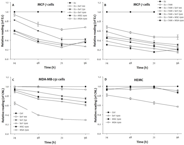
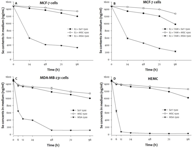
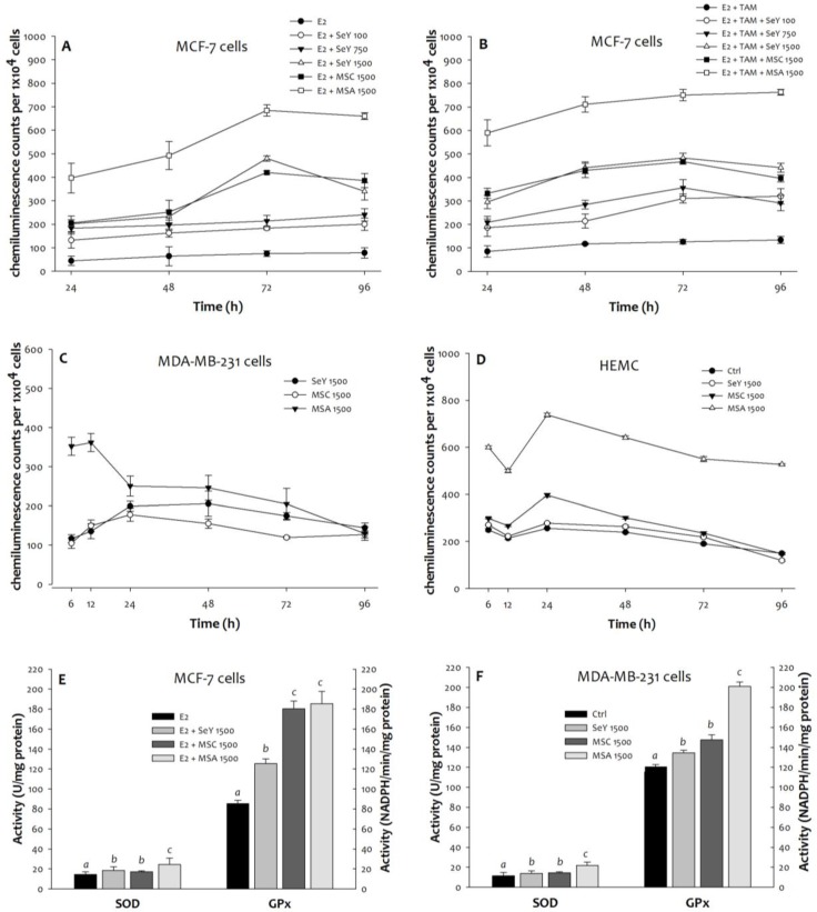
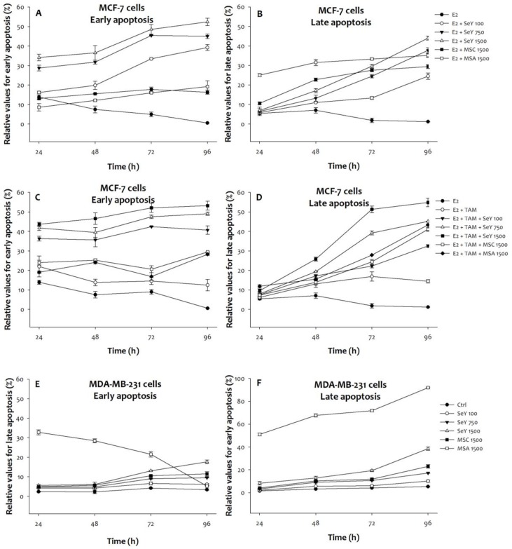
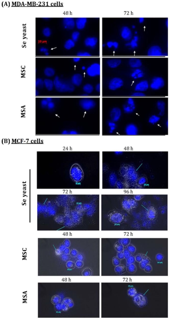
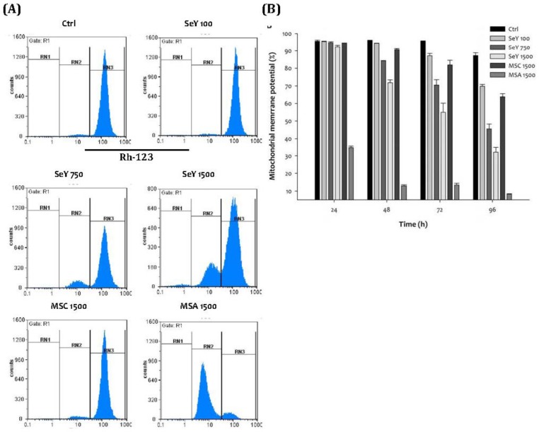
Recent evidence suggests that selenium (Se) yeast may exhibit potential anti-cancer properties; whereas the precise mechanisms remain unknown. The present study was aimed at evaluating the effects of Se yeast on oxidative stress, growth inhibition, and apoptosis in human breast cancer cells. Treatments of ER-positive MCF-7 and triple-negative MDA-MB-231 cells with Se yeast (100, 750, and 1500 ng Se/mL), methylseleninic acid (MSA, 1500 ng Se/mL), or methylselenocysteine (MSC, 1500 ng Se/mL) at a time course experiment (at 24, 48, 72, and 96 h) were analyzed. Se yeast inhibited the growth of these cancer cells in a dose- and time-dependent manner. Compared with the same level of MSA, cancer cells exposure to Se yeast exhibited a lower growth-inhibitory response. The latter has also lower superoxide production and reduced antioxidant enzyme activities. Furthermore, MSA (1500 ng Se/mL)-exposed non-tumorigenic human mammary epithelial cells (HMEC) have a significant growth inhibitory effect, but not Se yeast and MSC. Compared with MSA, Se yeast resulted in a greater increase in the early apoptosis in MCF-7 cells as well as a lower proportion of early and late apoptosis in MDA-MB-231 cells. In addition, nuclear morphological changes and loss of mitochondrial membrane potential were observed. In conclusion, a dose of 100 to 1500 ng Se/mL of Se yeast can increase oxidative stress, and stimulate growth inhibitory effects and apoptosis induction in breast cancer cell lines, but does not affect non-tumorigenic cells.

Keywords: Apoptosis; Breast Cancer Cells; Oxidative Stress; Selenium Yeast.


Conflict of interest statement

Competing Interests: The authors have declared that no competing interest exists.

Figures

Figure 1
Figure 1
Growth inhibition in (A)MCF-7 cells cultured with E2, (B)MCF-7 cells cultured with E2 and tamoxifen, (C)MDA-MB-231 cells, and (D)HEMC after 24, 48, 72, and 96 h incubation by Se yeast, MSC, and MSA. Results are expressed as relative reading (mean ± SD) at least from three independent experiments. Ctrl=controls; SeY 100=selenium yeast (100 ng Se/ml); SeY 750=selenium yeast (750 ng Se/ml); SeY 1500=selenium yeast (1500 ng Se/ml); MSC 1500=methylselenocysteine (1500 ng Se/ml); MSA 1500=methylseleninic acid (1500 ng Se/ml); E2=17β-estradiol; E2 + TAM=17β-estradiol and tamoxifen.
Figure 2
Figure 2
The Se levels of media was quantified in (A)MCF-7 cells cultured with E2, (B)MCF-7 cells cultured with E2 and tamoxifen, (C)MDA-MB-231 cells, and (D)HEMC after 0, 6, 12, 24, 48, 72, or 96 h incubation. Results are expressed as relative reading (mean ± SD) at least from three independent experiments. SeY 1500=selenium yeast (1500 ng Se/ml); MSC 1500=methylselenocysteine (1500 ng Se/ml); MSA 1500=methylseleninic acid (1500 ng Se/ml); E2=17β-estradiol; E2 + TAM=17β-estradiol and tamoxifen.
Figure 3
Figure 3
Superoxide generation in (A)MCF-7 cells cultured with E2, (B)MCF-7 cells cultured with E2 and tamoxifen, (C)MDA-MB-231 cells, and (D)HEMC after 6, 12, 24, 48, 72, and 96 h incubation, as well as (E)MCF-7 cells and (F)MDA-MB-231 cells after incubation of 48 h by Se yeast, MSC, and MSA. Results are expressed as relative reading (mean ± SD) at least from three independent experiments. SeY 1500=selenium yeast (1500 ng Se/ml); MSC 1500=methylselenocysteine (1500 ng Se/ml); MSA 1500=methylseleninic acid (1500 ng Se/ml).
Figure 4
Figure 4
Se compounds induced early and late apoptosis in (A)(B)MCF-7 cells cultured with E2, (C)(D)MCF-7 cells cultured with E2 and tamoxifen, and (E)(F) MDA-MB-231 cancer cells after 24, 48, 72, and 96 h incubation. Results are expressed as relative reading (mean ± SD) at least from three independent experiments. Ctrl=controls; SeY 100=selenium yeast (100 ng Se/ml); SeY 750=selenium yeast (750 ng Se/ml); SeY 1500=selenium yeast (1500 ng Se/ml); MSC 1500=methylselenocysteine (1500 ng Se/ml); MSA 1500=methylseleninic acid (1500 ng Se/ml); E2=17β-estradiol; E2 + TAM=17β-estradiol and tamoxifen.
Figure 5
Figure 5
Nuclear morphological changes induced by different Se compounds in (A)MDA-MB-231 cancer cells, as well as (B)MCF-7 cells cultured with E2 for different time periods. DAPI staining shows apoptotic nuclei; human breast cancer cells were exposure to different Se compounds (1500 ng Se/ml); MSC=methyl- selenocysteine; MSA=methylseleninic acid.
Figure 6
Figure 6
(A)Depolarization of mitochondrial membrane potential in MDA-MB-231 cells treated with Se compounds after 48 h incubation; (B)Mitochondrial membrane potential decrease caused by treated with Se compounds after 24, 48, 72 and 96 h incubation. Results are expressed as relative reading (mean ± SD) at least from three independent experiments. Ctrl=controls; SeY 100=selenium yeast (100 ng Se/ml); SeY 750=selenium yeast (750 ng Se/ml); SeY 1500=selenium yeast (1500 ng Se/ml); MSC 1500=methylselenocysteine (1500 ng Se/ml); MSA 1500=methylseleninic acid (1500 ng Se/ml).

References

    1. Herrera AC, Panis C, Victorino VJ, Campos FC, Colado-Simão AN, Cecchini AL, Cecchini R. Molecular subtype is determinant on inflammatory status and immunological profile from invasive breast cancer patients. Cancer Immunol Immunother. 2012;61:2193–201. - PMC - PubMed
    1. Schnitt SJ. Classification and prognosis of invasive breast cancer: from morphology to molecular taxonomy. Mod Pathol. 2010;23(Suppl 2):S60–4. - PubMed
    1. Guo CH, Hsia S, Chen PC. Distribution of selenium and oxidative stress in breast tumor-bearing mice. Nutrients. 2013;5:594–607. - PMC - PubMed
    1. Pani G, Galeotti T, Chiarugi P. Metastasis: cancer cell's escape from oxidative stress. Cancer Metastasis Rev. 2010;29:351–78. - PubMed
    1. Inokuma T, Haraguchi M, Fujita F, Tajima Y, Kanematsu T. Oxidative stress and tumor progression in colorectal cancer. Hepatogastroenterology. 2009;56:343–7. - PubMed

Publication types

MeSH terms